.webp&w=384&q=75)

Loading, please wait...
Loading, please wait...

Ovarian immature teratomas represent rare malignant germ cell neoplasms that predominantly affect adolescents and young women. Consequently, differentiating these aggressive lesions from benign mature dermoid cysts before surgery remains a formidable diagnostic challenge. Recent clinical research now highlights a distinct sonographic hallmark termed the pebbled mosaic sign to refine diagnostic accuracy and staging.
Clinicians frequently encounter diagnostic dilemmas when evaluating pelvic masses in adolescents. In particular, ovarian immature teratomas constitute less than one percent of all ovarian cancers. However, they account for up to twenty percent of malignant ovarian neoplasms in patients under twenty years of age. Therefore, early identification plays a pivotal role in optimizing oncologic outcomes. A recent retrospective cohort study at Peking Union Medical College Hospital evaluated thirty female patients with pure immature teratomas. The median patient age was 25.2 years, spanning an age range from 7 to 39 years. Furthermore, laboratory investigations revealed that 56.7% of these patients presented with elevated serum alpha-fetoprotein levels. This biochemical marker frequently alerts clinicians to an active malignant germ cell tumor. Additionally, most patients presented at early clinical stages. Specifically, FIGO stage IA accounted for 46.7% of cases, while stage IC comprised 50.0% of the cohort. The median maximum tumor diameter measured 16.7 centimeters, demonstrating the typically massive volume of these pelvic masses. As a result, comprehensive pelvic imaging remains essential to characterize internal architectural complexity before any surgical exploration.
Grayscale ultrasonography provides crucial insights into the internal architecture of these rare neoplasms. In this study, expert sonologists identified solid components in every single examined mass. Specifically, 30.0% of the tumors appeared purely solid, whereas 70.0% displayed a complex cystic-solid morphology. Most importantly, all thirty tumors exhibited a distinctive sonographic pattern known as the pebbled mosaic sign. Sonologists describe this hallmark as heterogeneous echogenicity situated on a hypoechoic grayscale background. Furthermore, the lesion features scattered punctate hyperechoic foci, localized fatty-like homogeneous hyperechoic zones, and tiny anechoic vesicles. Consequently, this unique acoustic texture resembles a pebbled mosaic or frothy pavement under high-resolution imaging. In contrast, benign mature teratomas typically demonstrate acoustic shadowing from dense calcifications or localized Rokitansky nodules. Thus, recognizing this mosaic texture allows clinicians to distinguish immature lesions from benign dermoids. Moreover, surgical teams observed spontaneous or intraoperative tumor rupture in 77.8% of patients whose masses presented with purely solid morphology. Because capsule disruption alters staging from stage IA to IC, sonographic detection of purely solid features carries immense clinical relevance.
Color Doppler interrogation provides vital diagnostic data regarding intratumoral perfusion. In this cohort, moderate vascularity appeared in 76.7% of the evaluated tumors. Meanwhile, minimal vascularity occurred in 20.0% of lesions, and only 3.3% displayed abundant blood flow. Therefore, immature teratomas often present with low to moderate vascularity, occasionally misleading clinicians into suspecting benign disease. Nevertheless, applying standardized scoring systems prevents underestimation of malignancy risk. Specifically, clinicians utilize the O-RADS classification framework to standardize ovarian mass risk assessment. Because all study lesions contained sizable solid components with irregular architecture, sonologists classified these tumors into high-risk O-RADS categories. Furthermore, power Doppler examination frequently demonstrates tortuous peripheral vessels supplying the hypercellular solid elements. In addition, hemodynamic evaluation assists surgical oncologists in anticipating vascularity during dissection. Consequently, combining grayscale pattern recognition with vascular scoring ensures comprehensive risk stratification. Radiologists should avoid categorizing large complex adnexal masses as benign dermoids when prominent vascular solid elements dominate the image. Thus, rigorous multiparametric sonography remains the cornerstone of preoperative germ cell tumor evaluation.
Pathological grading directly influences clinical prognosis and guides adjuvant chemotherapy protocols. Under microscopic examination, pathologists classify immature teratomas into grades 1, 2, or 3 based on the quantity of immature neuroepithelial tissue. In this study, histological evaluation revealed grade 1 in 36.7%, grade 2 in 36.7%, and grade 3 in 26.7% of patients. Importantly, statistical analyses uncovered a significant correlation between sonographic morphology and pathological grade. Specifically, tumors presenting with purely solid architecture, classified as pattern 1, correlated strongly with higher pathological grades. In contrast, tumors exhibiting larger cystic components and peripheral solid nodules more frequently corresponded to lower histological grades. Furthermore, Spearman rank correlation and Fisher exact testing verified this morphological association. Higher-grade lesions contain extensive primitive neuroectodermal tubules and cellular sheets that reflect sound waves differently than mature cystic cavities. As a result, a predominantly solid mass on ultrasound warrants heightened concern for grade 2 or grade 3 disease. Therefore, preoperative sonographic grading provides critical preliminary intelligence before final histological results become available. Hence, sonologists and pathologists must communicate closely when evaluating these heterogeneous specimens.
Accurate preoperative characterization holds profound ramifications for young female patients. Because immature teratomas predominantly arise in reproductive-age women, gynecologic oncologists prioritize fertility preservation whenever oncologically safe. Unilateral salpingo-oophorectomy with thorough surgical staging represents the standard of care for apparent stage I disease. However, unintended tumor rupture significantly increases the risk of peritoneal dissemination and elevates the disease stage to IC. As this study revealed, 77.8% of patients with purely solid tumors experienced rupture. Therefore, surgeons must exercise extreme caution during laparoscopic or open manipulation of large solid masses. In addition, identifying high-risk sonographic patterns alerts the surgical team to prepare for comprehensive staging, including peritoneal washings and omental biopsies. Moreover, high-grade tumors typically mandate platinum-based adjuvant chemotherapy following surgery, whereas stage IA grade 1 tumors may undergo surveillance alone. Consequently, preoperative identification of the pebbled mosaic sign empowers clinicians to plan fertility-sparing procedures while counseling families appropriately. Multidisciplinary tumor boards benefit immensely from integrating detailed sonographic pattern analysis into clinical decision-making. Ultimately, early and meticulous imaging assessment safeguards both oncologic survival and future reproductive potential.
The pebbled mosaic sign describes a characteristic heterogeneous sonographic pattern within the solid portions of immature teratomas. Sonologists observe a hypoechoic grayscale background embedded with punctate hyperechoic foci, fatty hyperechoic areas, and small anechoic microcysts. Consequently, this architectural arrangement produces a distinctive mosaic or cobblestone appearance. Recognizing this feature helps differentiate malignant immature teratomas from benign mature cystic teratomas, which usually demonstrate prominent Rokitansky nodules with dense acoustic shadowing.
Purely solid tumor morphology on ultrasound correlates strongly with higher histological grades in immature teratomas. Pathologists grade these tumors according to the quantity of immature neuroectodermal tissue present under microscopy. High-grade tumors, classified as grade 2 or 3, contain extensive sheets of primitive neuroepithelium that manifest sonographically as dense, solid masses. Conversely, tumors displaying prominent cystic components often represent low-grade disease, enabling clinicians to estimate biological aggressiveness before definitive pathological confirmation.
Preoperative ultrasound assessment guides surgical technique and safeguards fertility in young patients. Purely solid lesions carry a high risk of capsule rupture, which occurred in over seventy percent of solid cases. Intraoperative rupture spills malignant cells into the peritoneum, upstaging the disease and necessitating systemic chemotherapy. Therefore, identifying suspicious features alerts surgeons to avoid aggressive traction, select an appropriate surgical approach, and perform meticulous fertility-sparing staging while maintaining oncologic safety.
Disclaimer: This content is for informational and educational purposes only... Refer to the latest local and national guidelines for clinical practice.
References

Read summarized clinical updates, watch expert medical content, and earn CME certifications right from your smartphone.
Open your smartphone camera to scan and install.





Ovarian immature teratomas display a distinct pebbled mosaic sign on ultrasound. Solid tumor morphology correlates with higher pathological grades and increased rupture risk, highlighting the vital role of sonographic pattern recognition in guiding fertility-sparing surgery.
Today
Researchers in Hebei, China identified two novel HIV-1 unique recombinant forms, BDNY017 and BDK025, among patients failing antiretroviral therapy. Genomic analysis revealed complex mosaic structures involving CRF07_BC, CRF08_BC, and subtype B, alongside key drug resistance mutations including M184V, A98G, and E138K.
Today

A nationwide study covering 3.7 million Swedish births reveals that prenatal exposure to TORCH pathogens significantly elevates long-term risks of intellectual disability and autism. Healthcare providers must recognize clinical indicators, promote prenatal prevention, and track early pediatric neurodevelopment.
Today
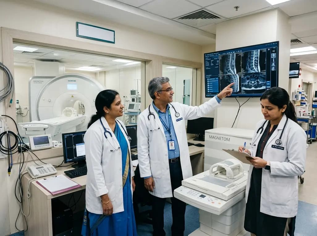
Under India's PLI initiative, domestic production of 57 critical medical devices has commenced, including MRI and CT scanners, cath labs, and heart valves. Global partnerships and local investments are expanding high-end technology, curbing import reliance, and strengthening clinical accessibility nationwide.
Today

A longitudinal study reveals that patients undergoing staged bilateral hip arthroscopy experience progressive contralateral labral deterioration during surgical intervals. However, structural degradation does not compromise 2-year postoperative clinical outcomes or patient-reported satisfaction scores.
Today

SWEDEPAD-1 trial insights show paclitaxel-coated devices do not improve long-term limb salvage in patients with chronic limb-threatening ischemia and tissue loss. While one-year reinterventions decreased, the devices were linked to a higher risk of major amputation at three months.
Today
