

Loading, please wait...
Loading, please wait...

Neoadjuvant chemotherapy plays a pivotal role in downsizing locally advanced tumors before surgical resection. However, conventional anatomical modalities struggle to differentiate viable residual cancer from treatment-induced fibrosis. Recent clinical investigations demonstrate that optoacoustic imaging breast cancer monitoring provides critical functional insights long before physical tumor shrinkage occurs. By coupling laser light with ultrasound detection, this hybrid modality tracks alterations in hemoglobin concentration and tissue oxygenation. Consequently, oncologists and radiologists gain an unprecedented noninvasive tool to tailor systemic regimens early during patient care.
Optoacoustic imaging integrates laser excitation with acoustic detection to generate high-contrast physiological maps. Specifically, short laser pulses irradiate breast tissue, inducing transient thermoelastic expansion of endogenous chromophores. Because hemoglobin absorbs light significantly more than surrounding parenchyma, acoustic detectors capture crisp ultrasonic waves from microvessels. This physiological mapping allows clinicians to evaluate tumor vascularity and blood oxygenation simultaneously in real time. In addition, the technique eliminates exposure to ionizing radiation, ensuring high patient safety during serial examinations.
Furthermore, malignant breast tumors depend heavily on abnormal angiogenesis to sustain rapid proliferation. These disorganized vessels exhibit irregular branching, heightened permeability, and fluctuating hypoxia. Traditional ultrasound depicts only physical mass boundaries, which change slowly across multiple treatment cycles. In contrast, optoacoustic platforms detect functional disruptions within tumor vasculature within days of initiating therapy. Consequently, sonologists observe vascular shutdown before tumor cells physically disappear. Therefore, monitoring hemodynamic shifts reliably distinguishes responsive lesions from chemoresistant disease long before macroscopic shrinkage manifests.
A recent prospective feasibility study analyzed twenty breast cancer patients undergoing neoadjuvant systemic chemotherapy through serial assessments. Researchers gathered imaging scans at baseline, after cycle one, at mid-treatment, and immediately prior to surgery. Notably, standard ultrasound volume measurements failed to correlate with pathologic complete response after cycle one. Similarly, volumetric shrinkage at mid-therapy did not distinguish complete responders from non-responders.
In contrast, functional optoacoustic parameters provided remarkable prognostic clarity after only one chemotherapy cycle. Specifically, patients who achieved a pathologic complete response demonstrated a significant decline in internal hemoglobin scores, falling to an average of seventy-six percent. Conversely, non-responders exhibited persistently high internal hemoglobin levels averaging nearly one hundred and nine percent. Thus, responsive tumors experienced prompt vascular collapse, whereas chemoresistant tumors maintained high vascular activity. Consequently, early internal hemoglobin reduction serves as a reliable surrogate marker of pathological eradication. Furthermore, clinicians can identify refractory disease after a single infusion, preventing weeks of ineffective exposure.
Accurate preoperative assessment dictates whether a surgical oncologist can safely perform breast-conserving therapy instead of mastectomy. In the feasibility study, all optoacoustic features decreased significantly more in complete responders than in non-responders before surgery. Specifically, evaluating internal hemoglobin, boundary zone vascularity, and peripheral vessels differentiated residual invasive malignancy from complete pathological resolution.
Moreover, chemotherapy-induced scar tissue frequently creates confounding acoustic shadows on standard grayscale ultrasound. Consequently, surgeons relying solely on anatomical boundaries might excise excessive healthy tissue or leave positive margins behind. Because optoacoustic imaging visualizes intravascular hemoglobin rather than dense fibrous tissue, it clarifies the true margins of residual disease. In addition, this functional precision assists surgical teams in de-escalating operative radicality whenever tumors resolve completely. Therefore, breast surgeons can design personalized conservation approaches with superior oncological confidence and improved cosmetic outcomes. Furthermore, real-time vascular mapping helps clinicians confirm clear margins intraoperatively, minimizing the need for secondary re-excision procedures.
Breast cancer remains the most frequently diagnosed malignancy among Indian women, often presenting at advanced stages. Because many patients present with palpable or locally advanced lumps, oncologists in India frequently utilize neoadjuvant chemotherapy to achieve surgical operability. However, peripheral healthcare facilities often rely solely on clinical palpation or basic ultrasound, which cannot detect early therapeutic failure.
Furthermore, contrast-enhanced dynamic magnetic resonance imaging imposes substantial financial burdens and remains unavailable across most rural cancer facilities. In contrast, handheld optoacoustic ultrasound offers a cost-effective, radiation-free alternative well-suited for Indian tertiary and district hospitals. Because the technology integrates directly into conventional ultrasound consoles, trained sonologists can easily incorporate hemodynamic scoring into routine follow-up visits. Additionally, identifying drug resistance after one cycle allows Indian oncologists to switch costly chemotherapy regimens promptly. Thus, timely regimen modifications reduce unnecessary financial toxicity and protect patients from pointless side effects. Ultimately, widespread deployment of functional ultrasound tools can democratize advanced response monitoring across resource-constrained Indian healthcare settings.
Translating optoacoustic biomarkers into standard oncology workflows requires robust scoring criteria and structured training. Radiologists must systematically evaluate five distinct parameters, including internal vessels, internal hemoglobin, deoxyhemoglobin, peripheral zone, and boundary zone. As clinicians adopt this validated scoring methodology, standardized reports will facilitate clearer communication across multidisciplinary tumor boards.
Moreover, future multi-center clinical trials must evaluate whether modifying chemotherapy based on early optoacoustic findings improves overall patient survival. For instance, non-responsive patients could switch to targeted therapies or immunotherapy before completing grueling treatment schedules. In addition, integrating artificial intelligence algorithms into optoacoustic consoles will soon automate vessel segmentation and oxygenation calculations. Ultimately, functional imaging shifts oncology from passive morphological observation toward dynamic, real-time therapy guidance. Therefore, clinical adoption of these optical biomarkers will accelerate personalized cancer therapy worldwide. Furthermore, prospective registry data will help regulatory bodies establish reimbursement frameworks, facilitating broader hospital adoption.
Standard breast ultrasound relies strictly on acoustic impedance differences to display tissue anatomy, echogenicity, and lesion dimensions. However, it cannot evaluate microvascular hemodynamics or oxygen saturation within the lesion. In contrast, optoacoustic imaging combines pulsed laser light with ultrasound detection to evaluate optical absorption of hemoglobin. Consequently, it generates real-time functional maps of tumor vascularity and hypoxia, enabling clinicians to assess biological response before anatomical shrinkage occurs.
Standard anatomical imaging cannot reliably differentiate between early responders and non-responders after a single treatment cycle. However, effective chemotherapy rapidly damages the fragile microvasculature feeding the tumor, precipitating an early decline in internal hemoglobin. In contrast, resistant tumors maintain robust vascular networks. Therefore, observing this vascular collapse after cycle one identifies patients heading toward complete pathological response, enabling medical oncologists to adjust regimens early for non-responding tumors.
Yes, optoacoustic imaging can help preserve breast tissue by providing superior discrimination between viable residual tumor and chemotherapy-induced fibrosis. Furthermore, post-treatment scar tissue often mimics persistent malignancy on conventional mammography and grayscale ultrasound. By demonstrating the complete absence of abnormal tumor hemoglobin and peripheral neovascularity, optoacoustic imaging confirms tumor eradication. Consequently, breast surgeons can confidently offer breast conservation surgery rather than performing extensive, unnecessary mastectomies.
Disclaimer: This content is for informational and educational purposes only and does not substitute professional medical advice, diagnosis, or treatment. Refer to the latest local and national guidelines for clinical practice.
References

Read summarized clinical updates, watch expert medical content, and earn CME certifications right from your smartphone.
Open your smartphone camera to scan and install.





A prospective feasibility study reveals that optoacoustic ultrasound imaging detects tumor hemoglobin reductions after cycle one of neoadjuvant therapy, predicting pathologic complete response earlier than conventional anatomical ultrasound in breast cancer.
Today
The Global Burden of Disease 2021 study reveals a dramatic rise in gastroesophageal reflux disease, with global cases surging over 80% since 1990. India carries the highest absolute burden worldwide, demanding intensified clinical screening, lifestyle interventions, and evidence-based therapeutic stewardship.
Today
A Swedish nationwide registry study of over 405,000 vaginal births shows that third-trimester SSRI exposure is linked to increased rates of labor induction, postpartum hemorrhage, and lower 5-minute Apgar scores, highlighting the need for balanced clinical risk-benefit counseling.
Today
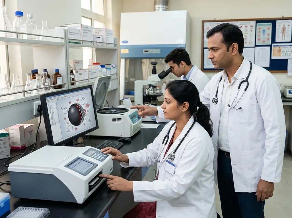
A novel MOF-based platform encapsulates hemoglobin with catalytic platinum and ceria nanozymes to deliver oxygen while scavenging toxic reactive oxygen species. This study highlights key design principles for managing hypoxia and reperfusion injury while resolving crucial stability challenges.
Today
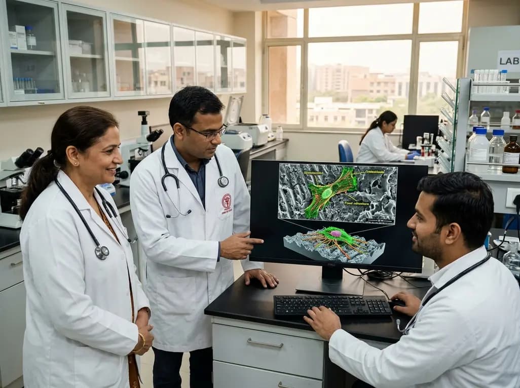
New research reveals that microscale topography on laser-textured Ti6Al4V governs osteoblast behavior and osseointegration regardless of high hydrophobicity. Dynamic interfacial evolution and controlled cytoskeletal tension enable robust cell adhesion, challenging traditional implant wettability determinism.
Today

A major World Health Organization study reveals that one in four teenagers globally records zero active days per week. Discover the epidemiological data, cardiometabolic consequences of adolescent sedentary behavior, and practical guidance for clinicians to promote healthy lifestyle habits among youth.
Today
