.webp&w=384&q=75)

Loading, please wait...
Loading, please wait...
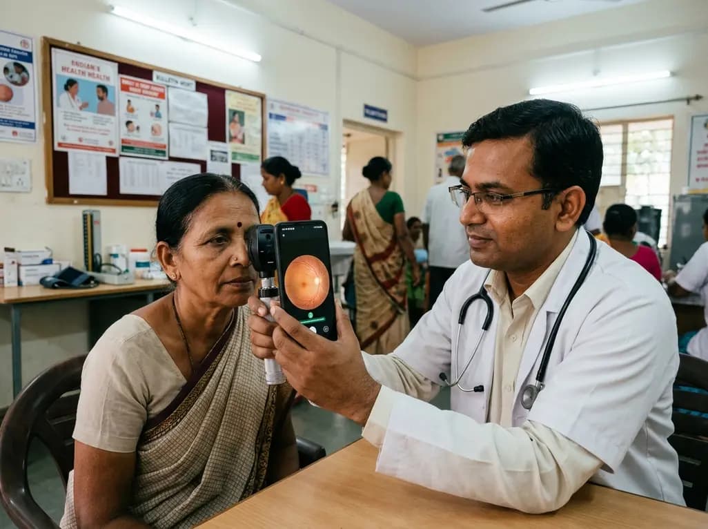
Modern ophthalmology faces a massive challenge in preventing avoidable blindness across underserved populations. Fortunately, recent advances in point-of-care diagnostic technology offer powerful new solutions. A landmark prospective study has demonstrated that offline AI retinal screening integrated into smartphone-based fundus cameras delivers exceptional diagnostic accuracy. Researchers at NIO Super Specialty Hospital in Pune evaluated the Medios-AI platform to assess its clinical utility. They discovered that this deep learning tool reliably identifies three leading causes of irreversible vision loss: diabetic retinopathy, glaucoma, and age-related macular degeneration. Consequently, clinicians can now execute sophisticated ophthalmic evaluations in decentralized community settings without relying on bulky infrastructure.
Historically, autonomous diagnostic platforms required substantial processing power and continuous cloud access. However, this innovative platform embeds complex neural networks directly onto handheld hardware. As a result, healthcare workers can capture high-resolution posterior pole images and obtain instant diagnostic reports at the bedside. The study, published in the prestigious European Journal of Ophthalmology, validates this smartphone-based approach in real-world cohorts. Therefore, this technological milestone represents a critical step forward for equitable healthcare delivery across India and other developing regions.
The clinical validation trial evaluated 371 eyes from 193 adult patients presenting with diverse ocular conditions or normal fundus anatomy. To establish a rigorous reference standard, two fellowship-trained vitreoretinal specialists graded widefield Clarus fundus photographs in a masked manner. Meanwhile, the offline AI system independently processed dilated fundus images captured through the Remidio Fundus on Phone device. Overall, the automated algorithm achieved a remarkable sensitivity of 99.3% and a specificity of 95.7% across the entire multi-disease cohort. These robust metrics prove that automated screening can match expert diagnostic precision in clinical practice.
Furthermore, disease-specific analysis demonstrated exceptional diagnostic performance across individual retinal conditions. The algorithm exhibited 98.2% sensitivity and 99.0% specificity for detecting glaucomatous optic neuropathy. For age-related macular degeneration, the system recorded 88.9% sensitivity and 97.5% specificity. In diabetic retinopathy cases, the tool demonstrated 84.6% sensitivity and 99.0% specificity. In addition, the software cleverly defaults to a combined diagnostic category when pathological features overlap between conditions. Consequently, primary care providers receive highly reliable classifications that prevent missed referrals for sight-threatening pathology.
Traditional teleophthalmology programs often falter in rural environments due to erratic telecommunications infrastructure. Cloud-dependent AI solutions require rapid image uploads and uninterrupted data streams to generate diagnostic insights. In contrast, this handheld platform executes all neural network computations locally on the mobile processor. Because the device functions entirely offline, clinicians can conduct comprehensive screening camps in remote geographical locations without network latency. Thus, healthcare teams eliminate data transfer bottlenecks and reduce expensive telecommunication overhead.
Moreover, local on-device processing significantly enhances data privacy and patient confidentiality. Sensitive biometric and retinal health records remain securely stored within the local device environment during screening drives. Health workers can instantaneously share printed or digital diagnostic summaries with patients before they leave the community venue. In addition, rapid report generation facilitates immediate patient counseling and timely specialist appointments. Consequently, patient compliance with downstream tertiary referrals improves substantially, ensuring prompt clinical interventions before irreversible vision loss occurs.
Rigorous clinical validation must accompany medical software before physicians deploy artificial intelligence in daily patient workflows. Notably, the Medios-AI platform has secured formal regulatory approval from the Central Drugs Standard Control Organisation in India for clinical deployment. Furthermore, the system carries European Class II medical device certification, confirming strict compliance with international safety and performance standards. These dual regulatory milestones give healthcare practitioners immense confidence in the diagnostic reliability of the device.
Primary care physicians, endocrinologists, and optometrists can now seamlessly incorporate autonomous retinal screening into their routine outpatient practices. For example, diabetic clinics can screen patients for diabetic retinopathy during regular metabolic checkups without dispatching them to separate diagnostic facilities. Similarly, general practitioners managing elderly individuals can rapidly identify early biomarkers of macular degeneration or glaucoma. Therefore, integrating validated smartphone diagnostic tools decentralizes essential ophthalmic care and bridges the severe specialist shortage in developing healthcare ecosystems.
Although these clinical results remain highly promising, physicians must recognize specific operational boundaries within current algorithms. As Dr. Aditya Kelkar observed, the present multi-disease algorithm is strictly validated for diabetic retinopathy, glaucoma, and age-related macular degeneration. Consequently, the tool cannot autonomously diagnose other vascular or inflammatory vitreoretinal disorders without dedicated algorithmic training. Each additional pathology demands separate prospective training datasets, independent clinical trials, and distinct regulatory clearances before commercial release.
Additionally, successful automated analysis depends heavily on adequate fundus image quality and pupillary clarity. Media opacities such as dense senile cataracts or corneal scars can impede adequate image capture, requiring specialist intervention. Furthermore, autonomous AI serves as a powerful triaging instrument rather than a complete substitute for comprehensive clinical evaluation. Therefore, clinicians must maintain established referral pathways so that identified suspects receive timely slit-lamp biomicroscopy, optical coherence tomography, and expert therapeutic management.
Deploying smartphone-based diagnostic systems offers a highly scalable model for public health screening initiatives. Paramedical staff and community health workers can master image acquisition protocols after brief standardized training sessions. Because the device provides real-time feedback on image gradability, operators can quickly recapture inadequate frames at the point of examination. Consequently, large-scale vision screening camps achieve superior throughput while maintaining stringent diagnostic accuracy standards.
Moreover, the integration of portable fundus cameras reduces capital expenditure compared to traditional tabletop fundus imaging equipment. District health authorities can equip primary health centers and mobile vision vans with cost-effective diagnostic kits. As a result, health systems maximize community outreach and alleviate the diagnostic burden on tertiary academic medical centers. Ultimately, adopting decentralized offline artificial intelligence establishes a sustainable framework for eliminating preventable blindness across vulnerable communities nationwide.
Q1: How does the smartphone-based offline AI system evaluate retinal images without internet connectivity?
The Medios-AI system embeds optimized deep learning neural networks directly into the smartphone fundus camera hardware. The internal mobile processor executes complex image processing and pattern recognition algorithms locally. Consequently, the device analyzes high-resolution fundus photographs in real time, generating comprehensive disease reports instantly at the point of care without requiring cloud servers or wireless networks.
Q2: Which specific ocular diseases can this smartphone AI algorithm accurately identify in patients?
The algorithm is clinically validated and approved to identify three major vision-threatening conditions: diabetic retinopathy, glaucoma, and age-related macular degeneration. In clinical trials, the system demonstrated an overall sensitivity of 99.3% and specificity of 95.7%. However, the system cannot currently diagnose other vitreoretinal conditions without further independent algorithm training and regulatory validation.
Q3: What regulatory approvals has this offline AI retinal screening technology received for clinical deployment?
The Medios-AI platform has received formal clinical approval from the Central Drugs Standard Control Organisation (CDSCO) in India. Additionally, the device holds European Class II medical device certification. These certifications validate its diagnostic accuracy, safety, and manufacturing compliance, allowing primary care physicians, optometrists, and eye hospitals to deploy the system safely in routine clinical practice.
Disclaimer: This content is for informational and educational purposes only. It does not constitute medical advice or replace professional judgment. Refer to the latest local and national guidelines for clinical practice.
References

Read summarized clinical updates, watch expert medical content, and earn CME certifications right from your smartphone.
Open your smartphone camera to scan and install.





A breakthrough study validates offline AI retinal screening integrated into smartphone fundus cameras, detecting diabetic retinopathy, glaucoma, and AMD with over 99% sensitivity. This point-of-care solution offers accurate, accessible, and cloud-independent eye care diagnostics for remote and underserved populations.
last month
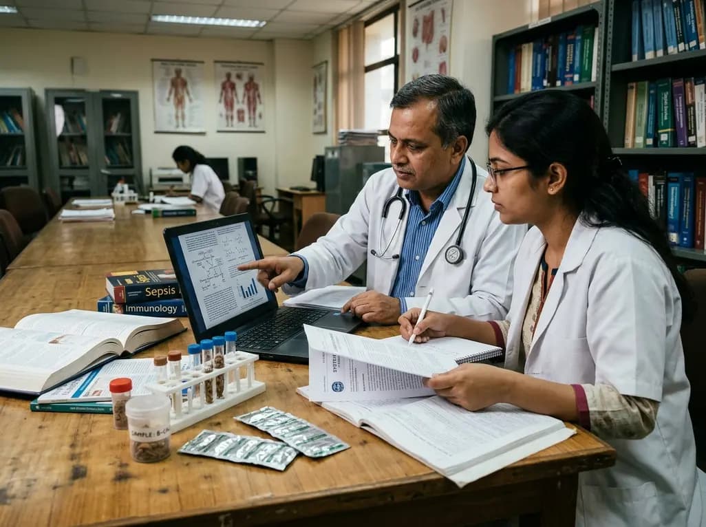
A systematic review of 41 preclinical studies reveals that baicalin dampens systemic inflammation, inhibits NF-κB and NLRP3 pathways, and improves organ preservation in sepsis models. However, substantial study heterogeneity and a complete lack of human clinical trials highlight the need for further translational research.
Today

Discover the crucial distinctions between mesonephric and mesonephric-like proliferations of the female genital tract. Learn the key histomorphologic, immunohistochemical, and molecular differences essential for accurate diagnosis and clinical management.
Today

A large UK Biobank study reveals that plasma proteomic signatures capture preclinical organ damage and improve multiorgan risk prediction across early CKM syndrome stages 0 to 2 beyond traditional PREVENT clinical models.
Today

A narrative review investigates whether the articularis genus muscle functions as an independent anatomical entity or blends with the vastus intermedius. We evaluate its morphology, role in retracting the suprapatellar bursa, and direct clinical significance in anterior knee pain and arthroplasty.
Today

A Bayesian multilevel meta-analysis reveals that aerobic training combined with moderate carbohydrate restriction modestly lowers HbA1c in type 2 diabetes. However, sparse data and very low certainty leave incremental benefits over exercise or diet alone unproven, highlighting the need for individualized care.
Today
