.webp&w=384&q=75)

Loading, please wait...
Loading, please wait...
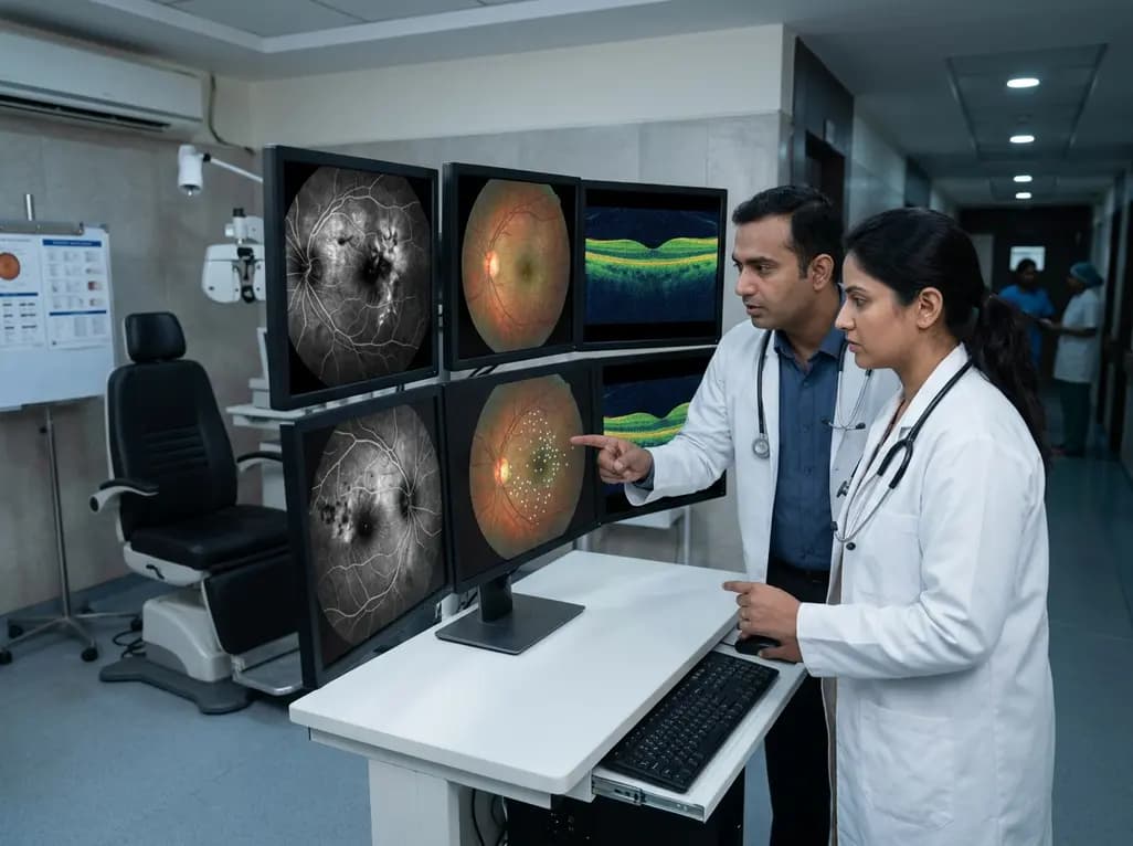
Chronic vascular macular oedema remains a leading cause of vision impairment in patients suffering from diabetic retinopathy and retinal vein occlusion. In India, where the burden of diabetic eye disease is exceptionally high, clinicians constantly seek precise diagnostic methods to improve treatment outcomes. A significant challenge in managing these cases involves the identification of retinal telangiectatic capillaries (TelCaps), which are often responsible for persistent fluid leakage. Traditional therapies, such as intravitreal anti-VEGF injections, sometimes yield a suboptimal response when these specific microvascular abnormalities are present. Consequently, retinal telangiectatic capillaries localization becomes crucial for planning targeted interventions like focal laser photocoagulation. Historically, indocyanine green angiography (ICG-A) has served as the reference standard for this task. However, the invasive nature of dye-based imaging often limits its repeatability and patient comfort. Recent advancements in optical coherence tomography (OCT) now offer a non-invasive alternative. By utilizing en face imaging techniques, ophthalmologists can potentially map these lesions with high precision without the risks associated with intravenous dyes. This transition toward structural imaging marks a significant shift in retinal diagnostics, promising more accessible and frequent monitoring for patients with chronic vascular diseases.
Retinal telangiectatic capillaries represent a distinct class of microvascular abnormalities that differ significantly from standard microaneurysms in their structural and functional profile. These lesions frequently appear as dilated, tortuous capillary segments that exhibit chronic leakage and contribute to the formation of hard exudates and persistent intraretinal fluid. In the context of retinal telangiectatic capillaries localization, understanding their behavior is essential for effective management. These TelCaps often cluster in areas of chronic ischemia or adjacent to circinate rings of exudates, indicating a high level of vascular permeability. Specifically, in Indian clinical settings, where patients may present with advanced diabetic maculopathy, these clusters can be particularly challenging to treat. While intravitreal therapies address the biochemical drivers of oedema, they may not always resolve the mechanical leakage from these structurally altered vessels. Therefore, localized focal laser photocoagulation remains a valuable adjuvant therapy. By targeting the source of the leakage directly, clinicians can achieve more durable stabilization of the retina. This targeted approach necessitates an imaging modality that can pinpoint the exact coordinates of the TelCaps within the various retinal layers, ensuring that the laser energy is applied effectively while minimizing damage to the surrounding healthy tissue.
Indocyanine green angiography has long been favored for imaging the deeper retinal and choroidal vasculature because its infrared properties allow it to penetrate through exudates and pigment. However, the study by Unger et al. demonstrates that structural OCT can achieve remarkable accuracy in retinal telangiectatic capillaries localization by leveraging en face reconstructions. The researchers analyzed images from 62 patients, comparing the performance of OCT cube B-scans against the traditional ICG-A reference. Notably, OCT identified 157 lesions compared to the 148 detected by ICG-A, suggesting that OCT might even capture certain structural changes that the dye-based method misses. The sensitivity for en face localized lesions reached 87%, with a positive predictive value of 82%. This high level of correlation indicates that structural OCT is not merely a supplementary tool but a viable primary imaging strategy for many patients. Furthermore, the ability to co-register OCT findings with infrared en face images provides a clear roadmap for surgeons. This spatial accuracy is particularly beneficial when TelCaps are located near the fovea, where surgical precision is paramount to avoid unintended scotomas. As a result, many centers may find that they can reduce their reliance on systematic ICG-A, reserving it for only the most complex cases.
The accuracy of OCT for retinal telangiectatic capillaries localization is significantly influenced by the surrounding retinal environment. According to the study findings, OCT performs exceptionally well when TelCaps are isolated or located adjacent to hard exudates. For isolated lesions, the sensitivity peaked at 92%, likely because the absence of surrounding vascular clutter allows for clearer visualization on B-scans. Conversely, the sensitivity dropped to 62% for clustered lesions. These clusters often present as a complex meshwork of microvascular abnormalities that can be difficult to resolve using purely structural criteria. Additionally, the presence of retinal thickening and exudates actually improved detection rates, as these features often serve as clinical markers that guide the investigator's focus. In contrast, false positives were more common in areas without significant retinal thickening. This suggest that the clinical context remains vital when interpreting OCT images. In Indian tertiary eye care centers, where patient volume is high, recognizing these nuances allows for better triage of imaging resources. While OCT excels in identifying well-defined, isolated TelCaps, clinicians must remain cautious when dealing with dense clusters where ICG-A might still provide superior contrast and resolution of the individual leaking points.
The move toward OCT-guided focal laser photocoagulation represents a modern evolution in the treatment of chronic macular oedema. Traditionally, surgeons relied on fluorescein or ICG angiography to guide their laser aim, which required alternating between different imaging platforms and sometimes dealing with outdated maps due to the time lag between angiography and the procedure. By utilizing en face OCT, clinicians can perform retinal telangiectatic capillaries localization using the same device used for routine monitoring. This integration streamlines the clinical workflow and allows for real-time planning. Moreover, because OCT is non-invasive, it can be repeated as often as necessary to track the resolution of TelCaps following treatment. In India, where cost-effectiveness and patient throughput are critical, reducing the need for expensive and time-consuming dye-based procedures can significantly improve healthcare delivery. However, the study emphasizes that ICG-A remains essential for complex, equivocal cases. Specifically, when OCT fails to provide a clear distinction between various microvascular changes, the dynamic flow information provided by angiography becomes indispensable. Therefore, a multimodal approach, starting with high-resolution OCT and escalating to ICG-A only when necessary, currently offers the most robust clinical strategy for managing these difficult-to-treat retinal pathologies.
The study by Unger et al. found that OCT has a high sensitivity of approximately 87% for localizing these microvascular abnormalities compared to the ICG-A gold standard. This high performance suggests that structural OCT is a reliable alternative for most patients. However, the sensitivity varies based on the lesion type, being much higher for isolated capillaries than for complex, clustered microvascular abnormalities.
Hard exudates often form a circinate pattern around leaking telangiectatic capillaries, serving as a biological marker for the underlying pathology. Clinicians can use these exudates to narrow their search area on OCT cube scans. The structural changes in the retina associated with these exudates, such as localized thickening, provide additional clues that enhance the detection and localization of the culprit leaking vessels.
While OCT is highly effective for most cases, ICG angiography remains essential for complex or equivocal scenarios, particularly when lesions are clustered together. Angiography provides dynamic information about blood flow and leakage that structural OCT cannot replicate. If the en face OCT images are unclear or if the patient is not responding to OCT-guided therapy, ICG-A should be utilized to gain a definitive map.
Disclaimer: This content is for informational and educational purposes only and does not constitute medical advice. It is not intended to be a substitute for professional medical judgment, diagnosis, or treatment. Always seek the advice of your physician or other qualified health provider with any questions you may have regarding a medical condition. Refer to the latest local and national guidelines for clinical practice.
References
Unger L et al. En face localization of retinal telangiectatic capillaries using OCT compared with ICG angiography in chronic vascular macular oedema. Acta Ophthalmol. 2026 Jul 03. doi: 10.1111/aos.70182. PMID: 42397689.
Hirano Y et al. Microvascular Abnormalities on Optical Coherence Tomography Angiography in Macular Edema Associated With Branch Retinal Vein Occlusion. Am J Ophthalmol. 2016;161:111-117.e1.
Shin JY et al. Optical coherence tomography-guided selective focal laser photocoagulation: a novel laser protocol for diabetic macular edema. Graefes Arch Clin Exp Ophthalmol. 2015;253(4):527-535.
"
Read summarized clinical updates, watch expert medical content, and earn CME certifications right from your smartphone.
Open your smartphone camera to scan and install.





A comparative study by Unger et al. evaluates the accuracy of non-invasive en face OCT versus ICG angiography for identifying retinal telangiectatic capillaries, offering a new pathway for guiding focal laser photocoagulation in chronic macular oedema.
3 months ago

Novel diimide and hydrazone scaffolds, particularly compound #C4, synergistically reactivate latent HIV-1 when combined with JQ1 or PKC activators. Demonstrating donor-matched proviral transcription ex vivo and non-canonical signaling, these findings advance shock and kill strategies toward reservoir eradication.
Today

A cross-sectional study reveals that maternal RSV vaccination coverage reached only 43.7% in Argentina, with maternal vaccine knowledge being the sole independent predictor of uptake. Clinicians play a crucial role in eliminating informational and structural barriers to optimize infant health.
Today

Review how tailored exercise programs benefit frail older patients with heart failure. Explore pathophysiological mechanisms, individualized training prescriptions, and strategies to overcome adherence barriers in clinical practice.
Yesterday

This comparative study evaluates posterior labral tears with and without glenohumeral instability, highlighting distinct demographic patterns, MRI underestimation in stable shoulders, and successful arthroscopic repair outcomes.
Yesterday

A clinical study demonstrates that early intervention with tocilizumab, either as monotherapy or bridged with corticosteroids, achieves significant proptosis reduction and resolves active inflammation in treatment-naive thyroid eye disease.
Yesterday
