.webp&w=384&q=75)

Loading, please wait...
Loading, please wait...

Malnutrition represents a pervasive yet frequently underestimated complication in patients with Crohn's disease and ulcerative colitis. Although clinicians routinely calculate body mass index during routine clinic visits, standard weight metrics often fail to capture critical shifts in lean muscle and hydration. Consequently, comprehensive nutritional assessment in IBD requires advanced diagnostic modalities that evaluate qualitative body composition. A prospective cohort study published in Inflammatory Bowel Diseases provides compelling evidence regarding how disease phase and care settings dictate body composition recovery and metabolic demands.
Traditional anthropometric measurements such as body mass index cannot differentiate between skeletal muscle loss, visceral adiposity, and fluid shifts. In active inflammatory bowel disease, systemic inflammatory cascades and catabolic cytokines trigger muscle wasting even when total body weight remains stable. Therefore, bioelectrical impedance vector analysis offers a reliable, non-invasive method to evaluate muscle mass and hydration without relying on standard population equations.
By utilizing bioelectrical impedance vector analysis, clinicians can calculate the fat-free mass index with greater accuracy. This metric objectively identifies hidden sarcopenia and subclinical malnutrition in both hospitalized individuals and outpatients. Furthermore, tracking vector shifts allows care teams to distinguish true cellular mass recovery from fluid retention caused by corticosteroid therapy or intravenous fluid administration. As a result, objective vector analysis provides clinicians with actionable diagnostic data that routine scale weight simply cannot deliver.
The prospective study conducted by Vrielink and colleagues investigated nutritional status and energy requirements across different phases of disease activity. Researchers monitored hospitalized patients with active flares, ambulatory outpatients with active disease, and patients in stable remission over a twelve-week longitudinal follow-up. They utilized indirect calorimetry to measure actual resting energy expenditure and bioelectrical impedance vector analysis to monitor lean mass changes.
Following clinical remission, hospitalized patients achieved a significant fat-free mass index increase of 1.1 kg/m². In contrast, ambulatory outpatients demonstrated a modest increase of only 0.4 kg/m², establishing a statistically significant difference between cohorts. Interestingly, body mass index changes and reductions in resting energy expenditure did not differ significantly between the two groups. Once patients achieved remission, their overall caloric balance matched the levels observed in the baseline remission cohort, demonstrating metabolic stabilization across care settings.
Several underlying physiological and clinical factors explain why hospitalized patients experienced greater increases in fat-free mass index during clinical recovery. First, hospitalized individuals often present with more severe systemic inflammation, profound initial catabolism, and marked muscle depletion at baseline. Consequently, when therapeutic interventions successfully suppress bowel inflammation, these patients experience pronounced anabolic rebound and rapid protein repletion.
Second, inpatient care typically provides structured nutritional supervision, rapid escalation of medical therapy, and managed caloric delivery. In contrast, ambulatory patients frequently struggle with self-imposed dietary restrictions, postprandial discomfort, and ongoing malabsorption at home. Furthermore, outpatients often maintain higher daily physical activity demands while enduring lingering subclinical gut inflammation. Therefore, structured clinical monitoring during acute hospital admissions may facilitate more robust muscle recovery compared to routine outpatient self-management strategies.
International clinical guidance from the European Society for Clinical Nutrition and Metabolism emphasizes early screening for malnutrition across all disease stages. Active inflammation accelerates systemic protein catabolism, alters gut permeability, and impairs nutrient absorption. Therefore, gastroenterologists and dietitians must not assume that normal or elevated body mass index excludes nutritional risk, particularly given the rising prevalence of sarcopenic obesity.
Clinical guidelines strongly advocate measuring resting energy expenditure and assessing body composition whenever feasible in complex cases. In addition, routine laboratory screening should evaluate micronutrient deficiencies, including iron, vitamin D, vitamin B12, and zinc. When indirect calorimetry is unavailable, clinicians should apply validated predictive energy equations while ensuring adequate protein provision between 1.2 and 1.5 grams per kilogram daily during active flares. Early multidisciplinary collaboration remains paramount for preventing irreversible lean tissue degradation.
To optimize patient recovery, clinical teams should implement serial body composition evaluations rather than relying solely on sporadic scale weights. Clinicians should incorporate validated screening tools such as the Malnutrition Universal Screening Tool or disease-specific questionnaires during every outpatient encounter. Moreover, when bioelectrical impedance vector analysis indicates low fat-free mass, targeted oral nutritional supplements enriched with high-quality protein should be prescribed immediately.
Furthermore, post-discharge care protocols must include dedicated nutritional counseling to sustain the anabolic gains achieved during hospital stays. Outpatients require practical dietary guidance that prevents unnecessary food group exclusions while alleviating gastrointestinal symptoms. Clinicians should also encourage gradual, structured resistance exercise during sustained remission to stimulate skeletal muscle protein synthesis. By combining targeted dietary interventions, anti-inflammatory therapies, and objective body composition monitoring, care teams can significantly improve long-term functional outcomes.
Bioelectrical impedance vector analysis evaluates tissue resistance and reactance directly, providing accurate estimates of cellular health and hydration without relying on fixed mathematical equations. In inflammatory bowel disease, this method detects hidden muscle wasting and sarcopenia that routine body mass index measurements consistently miss. Consequently, it allows clinicians to monitor genuine muscle mass recovery throughout anti-inflammatory treatment.
Hospitalized patients typically experience severe initial catabolism, creating significant physiological potential for rapid lean mass recovery once inflammation subsides. Furthermore, inpatient settings ensure direct medical supervision, prompt symptom control, and reliable delivery of macro- and micronutrients. Outpatients, conversely, often experience unmonitored dietary restrictions and persistent mild malabsorption that slow down skeletal muscle restoration.
During active inflammatory bowel disease flares, international nutritional guidelines recommend a daily protein intake between 1.2 and 1.5 grams per kilogram of body weight. This elevated intake counteracts accelerated protein catabolism, supports mucosal healing, and prevents rapid skeletal muscle depletion. Clinicians should combine this protein target with adequate caloric support to ensure optimal nitrogen balance.
Disclaimer: This content is for informational and educational purposes only and does not constitute medical advice, diagnosis, or treatment. Healthcare professionals must exercise independent clinical judgment. Refer to the latest local and national guidelines for clinical practice.
References
Vrielink TCO et al. Comparative assessment of nutritional status and energy expenditure in relation to dietary intake in hospitalized and outpatient patients with inflammatory bowel disease across disease phases. Inflamm Bowel Dis. 2026 Aug 30. doi: undefined. PMID: 42669191.
Bischoff SC, Biesalski HK, Valentini L, et al. ESPEN guideline on Clinical Nutrition in inflammatory bowel disease. Clin Nutr. 2023;42(3):352-379.
Favale A, Fadda A, Melis G, et al. Optimizing Malnutrition Risk Detection in Inflammatory Bowel Disease: A Longitudinal Analysis of Serial Nutritional Screening Tools. Nutrients. 2026;18(2):234-245.

Read summarized clinical updates, watch expert medical content, and earn CME certifications right from your smartphone.
Open your smartphone camera to scan and install.





A new prospective study reveals that hospitalized IBD patients achieve greater gains in fat-free mass index during remission compared to outpatients, despite similar BMI and energy expenditure metrics. Clinicians must move beyond BMI to monitor lean mass recovery and caloric balance accurately.
4 weeks back

Health authorities in the Democratic Republic of the Congo report unprecedented mortality from the Bundibugyo virus epidemic. Clinicians worldwide must understand the virological profile, atypical hemorrhagic manifestations, early diagnostic protocols, and containment strategies for this evolving health threat.
Today

A nationwide South Korean study shows underreporting of smoking among women dropped from 57.4% to 36.4% between 2008 and 2021. However, over one-third of female smokers still conceal their habit, highlighting the critical role of cotinine verification in clinical risk assessment.
Today
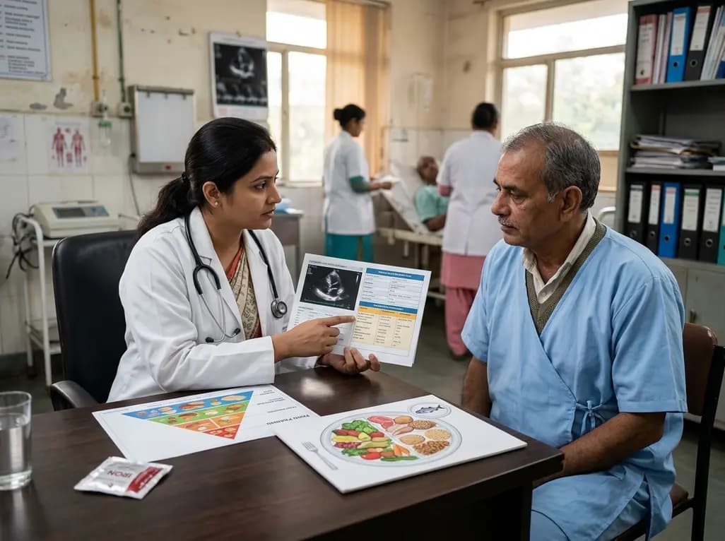
A scientific statement highlights the crucial role of nutrition in managing chronic heart failure. It emphasizes GLIM-based screening, individualized dietary patterns like the Mediterranean diet, appropriate protein and energy intake, avoiding unnecessary fluid restriction, and targeted iron supplementation.
Today

A new study evaluates the rheological stability of decellularized peripheral nerve matrix hydrogels under varied storage conditions. Findings show -80°C storage and freeze-drying preserve gelation kinetics and mechanical stiffness, offering key insights for shipping and translational clinical applications.
Today

A qualitative meta-synthesis highlights the complex interplay between gender dysphoria, minority stress, and eating disorders in transgender and gender-diverse individuals. Clinicians must recognize these distinct drivers to offer supportive, gender-affirming, and interdisciplinary eating disorder care.
Today
