.webp&w=384&q=75)

Loading, please wait...
Loading, please wait...
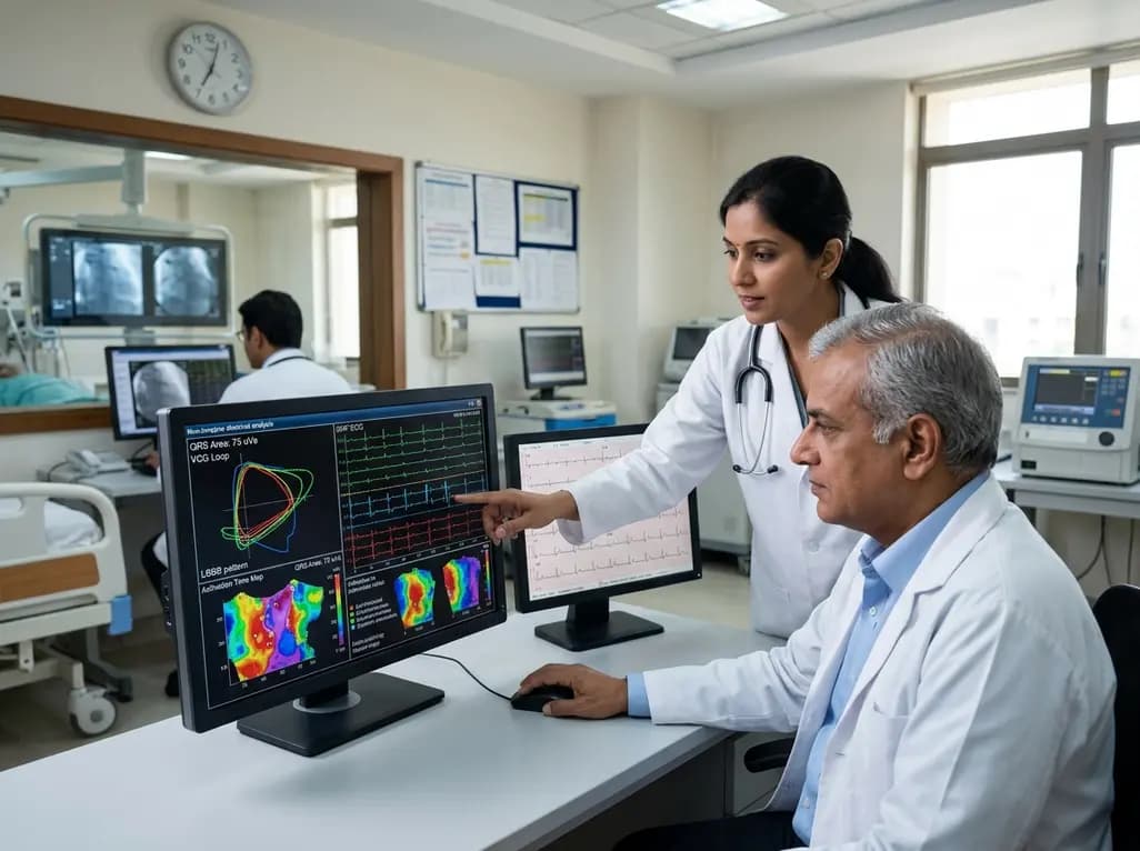
Heart failure management continues to advance as physiological pacing strategies challenge traditional biventricular resynchronization techniques. Recently, conduction system pacing CRT has emerged as an effective therapy to restore synchronized ventricular activation. However, not every patient achieves optimal electrical resynchronization after lead placement. Clinicians therefore require reliable, non-invasive modalities to identify prospective electrical responders before and during therapy delivery. A new investigation provides critical insights into non-invasive tools that accurately predict electrical recovery in heart failure patients.
Cardiac resynchronization therapy significantly improves functional capacity and reduces mortality in patients with heart failure and ventricular conduction delays. Traditional biventricular pacing stimulates the left ventricular epicardium via coronary sinus tributaries. Consequently, this non-physiological approach can produce heterogeneous activation patterns and incomplete resynchronization. In contrast, conduction system pacing directly recruits native conduction pathways through His-bundle or left bundle branch area pacing. As a result, this strategy generates rapid, physiological wavefront propagation across both ventricles. Nevertheless, variable clinical responses remain a persistent challenge in electrophysiology. Some individuals fail to achieve adequate electrical correction because of distal conduction disease, extensive myocardial fibrosis, or non-specific intraventricular conduction delays. Therefore, identifying physiological responders before invasive interventions remains essential. Deploying targeted non-invasive evaluations helps electrophysiologists identify candidates who will derive the greatest benefit from physiological pacing strategies, thereby minimizing procedural futility and enhancing patient care.
Standard surface electrocardiography provides the baseline evaluation for patients considered for resynchronization interventions. Historically, clinicians have relied on QRS duration and conventional bundle branch block definitions to guide selection. Researchers introduced Strauss criteria to define true left bundle branch block more strictly by incorporating duration thresholds alongside mid-QRS notching in multiple leads. In this new investigation of conduction system pacing, researchers evaluated how effectively Strauss criteria predict electrical resynchronization. The analysis revealed that Strauss criteria positivity yielded a positive predictive value of 83 percent. However, the negative predictive value was only 55 percent. These findings indicate that while classic Strauss criteria identify many true responders, they may exclude individuals who could still benefit from physiological pacing. Furthermore, standard surface recordings often miss subtle spatial vectors and localized activation delays. Therefore, electrophysiologists must combine standard surface tracings with advanced electrophysiological markers to evaluate ventricular activation accurately.
Vectorcardiography synthesizes electrical cardiac activity into a three-dimensional spatial representation of ventricular depolarization. By analyzing orthogonal X, Y, and Z leads, vectorcardiography quantifies unopposed electrical forces through the QRS area metric. In patients with significant conduction delays, a large QRS area signifies substantial dyssynchrony amenable to resynchronization. In the recent study, a baseline vectorcardiographic QRS area threshold of 100 µVs demonstrated exceptional diagnostic performance. Specifically, this cutoff yielded a positive predictive value of 100 percent and a negative predictive value of 73 percent. These findings highlight QRS area as a highly reliable non-invasive marker for selecting physiological pacing candidates. Furthermore, QRS area reflects global electrical dyssynchrony far more accurately than standard one-dimensional QRS duration measurements. Consequently, incorporating vectorcardiographic analysis into pre-procedural workflows enables clinicians to predict meaningful electrical resynchronization with remarkable precision and consistency.
Ultra-high-frequency electrocardiography represents an innovative non-invasive modality that records high-frequency electrical signals across standard precordial chest leads. Unlike conventional electrocardiograms, this technology captures microvolt potentials that reflect localized activation timing within myocardial regions. By computing the electrical dyssynchrony parameter, known as e-DYS, ultra-high-frequency electrocardiography precisely measures ventricular activation delay. In the analyzed study, an e-DYS threshold of 50 ms provided a positive predictive value of 93 percent and a negative predictive value of 47 percent. When researchers evaluated a 60 ms threshold, the positive predictive value remained robust at 90 percent. These results demonstrate that marked baseline electrical dyssynchrony strongly predicts successful resynchronization via conduction pathways. Moreover, ultra-high-frequency recordings provide instantaneous feedback during lead deployment, allowing implanters to confirm capture of the specialized conduction system immediately and optimize lead placement in real time.
Alongside electrical mapping, evaluating structural substrate via cardiac magnetic resonance imaging remains crucial. Myocardial scar burden, particularly within septal and inferolateral walls, significantly influences pacing success. When extensive transmural fibrosis replaces viable myocardium, electrical wavefronts encounter anatomical barriers and conduction blocks. Consequently, even successful pacing lead capture cannot properly propagate depolarization across scarred left ventricular tissue. Integrating non-invasive mapping with magnetic resonance imaging allows electrophysiologists to distinguish between pure conduction system disease and permanent tissue necrosis. Thus, optimizing patient outcomes requires moving toward personalized, multi-parametric diagnostic evaluations. For patients with isolated proximal conduction blocks and preserved myocardial tissue, conduction system pacing provides exceptional resynchronization efficiency. In contrast, patients with extensive distal scarring may require optimized hybrid or biventricular approaches. Therefore, adopting a comprehensive pre-implantation workflow improves procedural success rates and optimizes long-term ventricular remodeling.
Conduction system pacing CRT directly stimulates the intrinsic conduction network through His-bundle or left bundle branch area pacing. Consequently, this method restores natural, rapid physiological activation throughout both ventricles. In contrast, conventional biventricular pacing stimulates myocardial tissue from the epicardial coronary sinus branches, which often results in slower, non-physiological wavefront propagation and highly variable clinical responses across different patient populations.
Vectorcardiographic QRS area measures electrical forces in three dimensions across orthogonal planes, capturing spatial dyssynchrony and delayed activation forces comprehensively. In contrast, standard QRS duration only provides a one-dimensional measurement that frequently overlooks regional activation abnormalities. Therefore, vectorcardiographic QRS area offers significantly higher diagnostic accuracy and predictive value when identifying electrical resynchronization in prospective heart failure candidates undergoing physiological pacing.
Ultra-high-frequency ECG captures high-frequency myocardial signals to quantify regional ventricular activation delays and electrical dyssynchrony in real time. During lead placement procedures, this non-invasive tool helps implanters verify immediate capture of the specialized conduction system. Furthermore, it allows clinical operators to confirm physiological resynchronization instantly, thereby optimizing lead positioning, reducing procedure duration, and minimizing overall radiation exposure during cardiac interventions.
Disclaimer: This content is for informational and educational purposes only and does not constitute medical advice. Healthcare professionals should rely on their clinical judgment and refer to the latest local and national guidelines for clinical practice.
References

Read summarized clinical updates, watch expert medical content, and earn CME certifications right from your smartphone.
Open your smartphone camera to scan and install.





A recent study evaluates non-invasive tools, including VCG QRS area and UHF-ECG, to predict electrical resynchronization in patients undergoing conduction system pacing CRT.
last month

A Bayesian multilevel meta-analysis reveals that aerobic training combined with moderate carbohydrate restriction modestly lowers HbA1c in type 2 diabetes. However, sparse data and very low certainty leave incremental benefits over exercise or diet alone unproven, highlighting the need for individualized care.
Today

Findings from Project Viva indicate that sustained postpartum weight retention, rather than excessive gestational weight gain alone, directly worsens women's mid-life cardiovascular health, degrading blood pressure and glycemic parameters decades after delivery.
Today

A large UK Biobank study reveals that plasma proteomic signatures capture preclinical organ damage and improve multiorgan risk prediction across early CKM syndrome stages 0 to 2 beyond traditional PREVENT clinical models.
Today

A retrospective CBCT study reveals that superior TMJ space asymmetry correlates moderately with occipital condyle-atlas-axis asymmetry in unilateral TMJ osteoarthritis, highlighting an imaging correlate between jaw biomechanics and upper cervical alignment rather than a standalone diagnostic marker.
Today

A cross-sectional investigation highlights the complex pathophysiological links between serum Klotho deficiency, secondary hyperaldosteronism, and vascular damage in CKD. Discover key insights into non-invasive vascular stiffness markers and targeted therapeutic pathways for cardiorenal protection.
Today
