.webp&w=384&q=75)

Loading, please wait...
Loading, please wait...

Chronic liver disease represents a major cause of global morbidity and mortality. Because persistent hepatic injury prompts progressive extracellular matrix accumulation, accurate assessment of liver fibrosis is essential for clinical management. While percutaneous liver biopsy historically served as the primary diagnostic standard, its invasiveness and sampling error limit routine use. Consequently, non-invasive imaging modalities have emerged as indispensable staging alternatives.
Persistent parenchymal inflammation stimulates hepatic stellate cells, which drive fibrogenesis and alter vascular architecture. As fibrous bands expand, patients encounter heightened risks of portal hypertension, hepatic decompensation, and hepatocellular carcinoma. Therefore, clinicians require accurate staging tools to identify advanced disease before irreversible decompensation occurs. Routine serological panels, such as the AST-to-platelet ratio index, provide accessible initial screening in outpatient clinics. Nevertheless, serum biomarkers lack sufficient diagnostic accuracy to distinguish intermediate fibrosis stages reliably. In contrast, non-invasive imaging measures tissue elasticity directly, offering physical metrics that correlate with collagen deposition. Furthermore, imaging examinations visualize macroscopic liver morphology, identifying signs of portal hypertension such as splenomegaly. Accordingly, international guidelines recommend non-invasive imaging to guide surveillance intervals and pharmacotherapy initiation. In addition, early risk stratification helps hepatologists allocate medical resources and plan surveillance endoscopy efficiently. Consequently, timely detection prevents hospital admissions related to liver failure. Serial evaluations also confirm fibrosis regression following therapy. Thus, imaging-based staging forms the cornerstone of modern liver disease management.
Vibration-controlled transient elastography represents the most widely utilized point-of-care tool for assessing liver stiffness. The device uses a mechanical piston to propagate low-frequency shear waves into the hepatic parenchyma. Specialized ultrasound pulses then track shear wave velocity, converting this measurement into kilopascals. Because shear waves propagate faster through stiffened matrix tissue, numerical values reflect fibrosis severity reliably. In addition, modern systems incorporate the controlled attenuation parameter, which quantifies concomitant hepatic steatosis in decibels per meter. Consequently, clinicians obtain rapid, dual-parameter evaluations during outpatient consultations. However, transient elastography has well-documented technical limitations. Specifically, the system lacks real-time B-mode imaging, which prevents anatomical visualization during measurement acquisition. Operators cannot actively avoid large blood vessels or focal parenchymal lesions. Furthermore, thoracic adipose tissue dampens acoustic energy, increasing technical failure rates in obese patients. Although specialized XL probes improve feasibility in obesity, severe chest wall adiposity still impairs performance. Similarly, perihepatic ascites blocks shear wave propagation completely. Clinicians must therefore recognize these limitations when interpreting ambiguous clinical readings.
Ultrasound shear wave elastography overcomes major limitations of transient elastography by integrating stiffness quantification directly into standard diagnostic ultrasound systems. The technology employs acoustic radiation force impulses to induce microscopic shear waves within targeted liver parenchyma. High-frame-rate tracking then computes shear wave velocity, generating quantitative elastograms superimposed on live B-mode images. Clinicians select between point shear wave elastography and two-dimensional shear wave elastography depending on clinical needs. Because operators sample tissue under direct ultrasound visualization, they reliably avoid large vessels, biliary structures, and focal lesions. Furthermore, this anatomical guidance allows successful evaluations in patients with moderate ascites, where fluid-free acoustic windows exist. However, shear wave elastography requires strict adherence to standardized acquisition protocols. For instance, excessive transducer pressure artificially elevates measured liver stiffness, creating false-positive diagnoses. Moreover, proprietary calculation algorithms vary considerably among ultrasound manufacturers, producing discrepant values across different vendor machines. Therefore, longitudinal disease monitoring requires consistent equipment utilization to prevent erroneous diagnostic interpretations. Radiology teams must apply vendor-specific cutoffs to ensure clinical accuracy.
Magnetic resonance elastography delivers the highest diagnostic accuracy, reproducibility, and technical success among contemporary imaging techniques. The modality uses an active acoustic driver to transmit continuous low-frequency vibrations into the abdominal cavity. Simultaneously, specialized phase-contrast pulse sequences image microscopic wave displacement across the liver volume. Automated computer algorithms invert these displacement patterns into quantitative elastograms that display shear stiffness in kilopascals. In contrast to ultrasound modalities that interrogate narrow acoustic windows, magnetic resonance elastography samples the entire liver volume. Consequently, the technique eliminates sampling errors caused by the heterogeneous fibrosis distribution common in steatotic liver disease. Furthermore, severe obesity and ascites do not impair acquisition feasibility, ensuring reliable evaluation across complex cohorts. However, equipment costs, scanner availability, and longer examination times limit broad clinical accessibility. In addition, severe hepatic iron overload causes profound T2* signal attenuation, which can preclude valid calculations. Claustrophobia and metallic implants also restrict patient eligibility. Despite these practical constraints, magnetic resonance elastography remains the reference standard for resolving indeterminate ultrasound assessments.
Optimal management of chronic liver disease requires comprehensive tissue characterization extending beyond isolated stiffness metrics. Modern magnetic resonance imaging integrates advanced sequences that evaluate steatosis and fibro-inflammatory activity within a single scanning session. In particular, magnetic resonance imaging proton-density fat fraction serves as the non-invasive reference for fat quantification. Unlike ultrasound attenuation parameters, proton-density fat fraction calculates precise volumetric fat percentages without confounding from inflammation. Furthermore, iron-corrected T1 mapping evaluates necroinflammatory disease activity effectively. Elevated corrected T1 values indicate ongoing inflammation and cellular edema, helping clinicians distinguish active steatohepatitis from stable fibrosis. Integrating these quantitative biomarkers into tiered care pathways improves patient stratification. Primary care clinicians use point-of-care transient elastography to rule out advanced disease efficiently. Patients with ambiguous results then undergo shear wave elastography in hospital ultrasound clinics. When diagnostic uncertainty persists or detailed phenotyping is necessary, teams escalate patients to magnetic resonance examinations. Moreover, clinicians must account for physiological confounders such as acute hepatitis or recent meals. Thus, tiered pathways optimize diagnostic precision while conserving resources.
Acute physiological factors frequently elevate liver stiffness independently of true histological fibrosis. For example, acute hepatitis flares increase cellular edema, yielding falsely elevated stiffness values. Furthermore, postprandial hepatic hyperemia, extrahepatic biliary obstruction, and systemic venous congestion from right-sided heart failure elevate parenchymal tension. Consequently, clinicians must ensure that patients fast for at least three hours and review hepatic biochemistry before establishing a definitive fibrosis diagnosis.
Clinicians should prioritize magnetic resonance elastography when ultrasound elastography yields indeterminate results or technical failures, which occur frequently in severe obesity or marked ascites. Furthermore, magnetic resonance elastography interrogates the entire liver volume, overcoming the sampling bias of focal ultrasound measurements. It also incorporates complementary sequences, including proton-density fat fraction and iron-corrected T1 mapping, delivering comprehensive tissue phenotyping when detailed staging is required for complex hepatology management.
Serial elastography provides a dependable, non-invasive method for tracking chronic liver disease progression and evaluating therapeutic responses over time. Effective antiviral therapy or sustained lifestyle modifications often decrease liver stiffness, indicating resolving inflammation and fibrosis regression. However, clinicians should ensure that follow-up scans utilize identical elastography equipment and vendor platforms whenever feasible. This technical consistency prevents inter-vendor calibration discrepancies and permits accurate interpretation of longitudinal tissue elasticity changes.
Disclaimer: This content is for informational and educational purposes only and should not be considered medical advice. Always consult a qualified healthcare provider for clinical decisions. The views expressed here do not necessarily reflect the official policy or position of any healthcare institution or regulatory body. Refer to the latest local and national guidelines for clinical practice.
References

Read summarized clinical updates, watch expert medical content, and earn CME certifications right from your smartphone.
Open your smartphone camera to scan and install.





Non-invasive imaging techniques have revolutionized liver fibrosis assessment. This clinical review evaluates vibration-controlled transient elastography, ultrasound shear wave elastography, and MR elastography to optimize clinical decision-making and longitudinal care pathways for chronic liver disease.
Today

A comprehensive meta-analysis of over 278,000 women demonstrates that maternal atopic dermatitis significantly increases risks of premature rupture of membranes and low birth weight. Discover essential clinical insights on disease mechanisms, serial antenatal growth monitoring, and safe therapeutic interventions.
Today

Over 60 international public health researchers have advised FSSAI to mandate front-of-pack warning labels for any packaged product high in even one critical nutrient: sugar, saturated fat, or sodium. This reform counters phased rollouts to accelerate consumer protection and address India's chronic disease burden.
Today
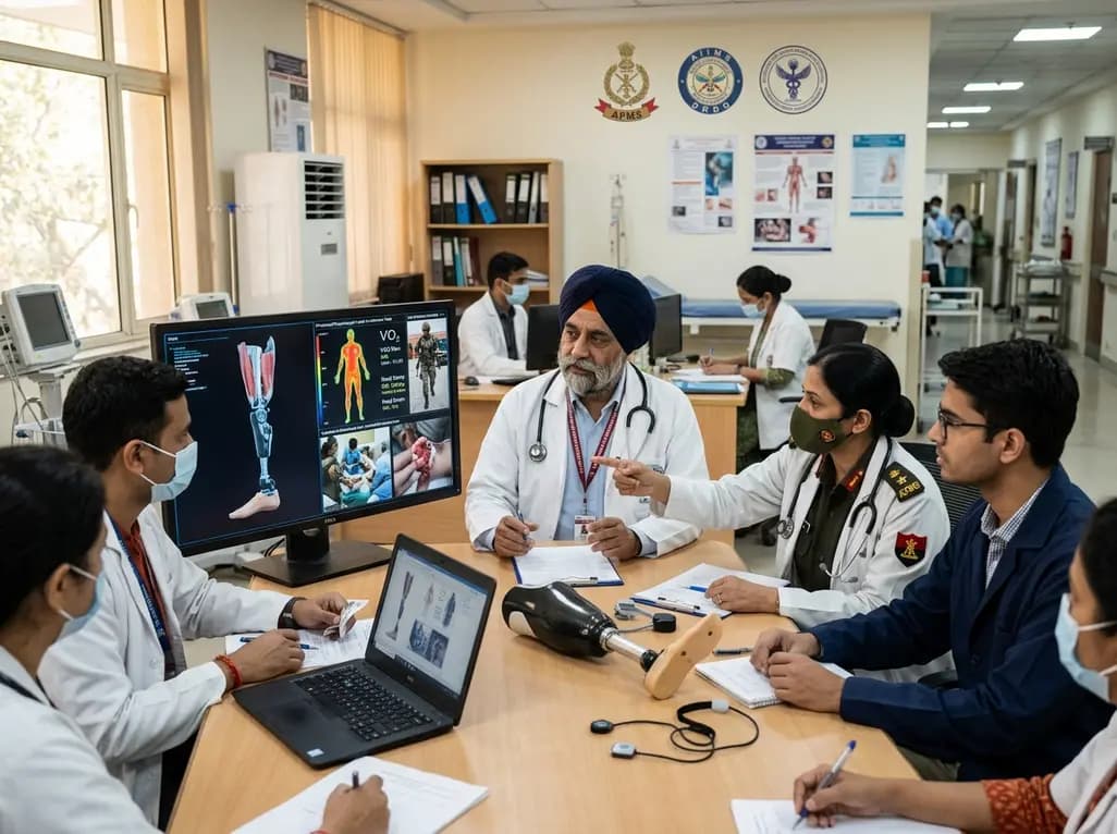
India's premier scientific bodies have joined the Armed Forces Medical Services to solve unique physiological and operational challenges faced by soldiers. The national collaboration covers combat casualty care, artificial intelligence diagnostics, bionic prosthetics, and extreme-environment physiological resilience.
Today

A cross-sectional study reveals that elevated METS-IR is independently associated with higher myopia odds in adolescents, showing biological synergy with obesity and highlighting the need for metabolic screening in pediatric eye care.
Yesterday
