.webp&w=384&q=75)

Loading, please wait...
Loading, please wait...

Deciphering how the human brain transforms perceptual experiences into durable memory remains a foundational challenge in modern neuroscience. Recent investigations provide groundbreaking insights into visual recognition memory by evaluating electroencephalographic signatures across learning and retrieval phases. Specifically, cognitive neuroscientists employed representational similarity analysis and multivariate cross-classification to decode complex neural activity. These computational techniques capture the evolving dynamics of visual processing across time. Therefore, researchers can track how initial perception converts into durable mnemonic codes. Historically, neuroscientists studied encoding and retrieval as isolated physiological phenomena. However, emerging empirical evidence indicates that these stages share coordinated electrophysiological dynamics. Healthy adults rapidly register sensory details before consolidating them into accessible representations. Consequently, precise temporal resolution from electroencephalography reveals early sensory discrimination alongside late retrieval signals. As a result, clinicians gain deeper clarity regarding how neural ensembles coordinate memory formation. Ultimately, understanding these shared signatures establishes an essential baseline for evaluating subtle cognitive decline in neurodegenerative illnesses.
Memory processing unfolds across precise millisecond milestones following stimulus exposure. Initially, the visual system categorizes visual stimuli within 150 milliseconds of stimulus onset. This early latency reflects rapid feedforward processing through primary and secondary visual cortices. Furthermore, distinct memory-related effects emerge across stimulus categories as early as 150 milliseconds post-onset. Notably, neural responses to encoding, previously encountered items, and novel stimuli begin diverging around 300 milliseconds. During this critical processing window, items presented during study phases exhibit pronounced differentiation from old items in testing sessions. Moreover, traditional old versus new electrophysiological effects consolidate strongly between 400 and 600 milliseconds. This prolonged latency corresponds to well-characterized parietal and frontal event-related potentials. Therefore, these late neural components reflect conscious episodic recollection and familiarity assessment. In contrast, earlier intervals reflect perceptual categorization. Consequently, the brain orchestrates a sequential continuum from sensory registration to mnemonic confirmation. In addition, multivariate cross-classification demonstrates that memory-specific patterns persist across varied visual objects. Thus, temporal chronometry reveals an integrated processing pipeline that systematically reorganizes perceptual inputs into lasting memories.
Visual stimulus categories do not engage cortical memory circuits in identical ways. In particular, human faces elicit distinct electrophysiological responses compared to ordinary inanimate objects. The study observed that short-term familiarization with arbitrary objects did not consistently mimic long-term personal familiarity. However, experimentally familiarized faces displayed statistically significant memory effects. Consequently, facial stimuli appear to recruit specialized neural pathways that accelerate mnemonic encoding. Evolutionary mechanisms have clearly prioritized human facial discrimination for social survival and communication. Therefore, the right fusiform gyrus and occipital face network operate with distinct temporal efficiency. Furthermore, anterior regions of interest, especially within the right hemisphere, did not differentiate study items from entirely novel items. This lack of differentiation indicates that the brain initially treats all newly presented stimuli through similar anterior processing channels. In contrast, familiar faces rapidly recruit distributed temporal networks. Moreover, these unique facial signatures emphasize the structural independence of socially relevant stimuli. Thus, evaluating face processing yields valuable diagnostic clues for identifying selective cortical lesions.
Traditional electrophysiological analyses often overlook subtle, multi-electrode pattern variations by focusing on isolated peak amplitudes. In contrast, multivariate pattern analysis provides a powerful framework to decode distributed brain activity across time. Researchers trained machine learning classifiers to distinguish visual stimulus categories and mnemonic status from multi-channel scalp data. Furthermore, cross-category classification demonstrated that memory retrieval shares underlying neural codes across divergent object domains. This algorithmic success proves that the brain forms abstract mnemonic representations beyond simple sensory features. Moreover, cross-dataset classification verifies that these electrophysiological patterns generalize reliably across different individuals and testing sessions. Consequently, multivariate methods bridge the gap between microscopic cellular firing and macroscopic scalp potentials. Additionally, representational similarity analysis clarifies how neural populations structure visual knowledge. Therefore, these neurocomputational techniques offer unprecedented sensitivity for probing high-level cognitive operations. Researchers can now identify subtle mnemonic disruptions before overt behavioral deficits emerge. Ultimately, multivariate classification establishes a rigorous foundation for modern cognitive electrophysiology and translational neuroscience.
Electrophysiological findings regarding visual memory offer profound practical implications for clinical neurology and psychiatry. For instance, amnestic mild cognitive impairment and early Alzheimer's disease typically compromise episodic retrieval pathways. Pathological amyloid deposition and tau neurofibrillary tangles preferentially destroy medial temporal lobe structures. Consequently, affected individuals struggle to consolidate novel visual information or distinguish old items from new distractors. By mapping the millisecond timing of visual recognition memory, electrophysiology uncovers early circuit failure. Furthermore, patients with frontotemporal lobar degeneration frequently display aberrant social cue processing and face recognition deficits. Disrupted 150-millisecond perceptual categorization can signify primary sensory cortical pathology. Meanwhile, delays in the 400 to 600 millisecond window reflect impaired hippocampal-cortical retrieval networks. Therefore, time-resolved electrophysiological testing can help clinicians differentiate cortical dementias from psychiatric disorders like pseudodementia. In addition, standardized cognitive neurophysiology enhances bedside clinical evaluation across Indian tertiary healthcare centers. Hence, characterizing shared memory signatures aids the objective assessment of progressive neurocognitive disorders.
Translating cognitive neuroscience into reliable medical biomarkers requires objective, reproducible analytical methodologies. Historically, clinicians relied on subjective neuropsychological questionnaires to gauge memory failure. However, cultural diversity, linguistic differences, and varying literacy levels across India frequently limit standard psychometric testing. In contrast, multivariate electroencephalographic markers provide objective, language-independent neurophysiological assessments of cognitive function. Because cross-classification algorithms identify shared neural representations, they deliver unbiased metrics of mnemonic integrity. Furthermore, these non-invasive electrophysiological biomarkers can track therapeutic efficacy in experimental drug trials. For example, neuroprotective interventions targeting synaptic preservation should theoretically rescue delayed 400-millisecond recognition dynamics. Additionally, combining time-resolved electroencephalography with structural neuroimaging provides comprehensive multimodal disease profiling. Physicians can monitor post-stroke cognitive recovery or traumatic brain injury rehabilitation with greater precision. Moreover, electrophysiology remains far more accessible than advanced functional neuroimaging. Consequently, widespread implementation across resource-conscious healthcare environments becomes feasible. Ultimately, integrating advanced computational electrophysiology into clinical pathways will enhance early diagnostic precision for vulnerable patients.
Visual recognition memory represents the cognitive capacity to identify previously encountered visual items or stimuli. This neurological process relies on coordinated activity across the occipitotemporal visual stream and medial temporal lobe structures. Consequently, intact recognition ensures proper distinction between novel environmental cues and familiar learned objects during daily human functioning.
Multivariate cross-classification decodes distributed patterns of electrical activity across scalp electrodes over precise millisecond intervals. Therefore, this computational approach tracks temporal representations without relying exclusively on single ERP amplitudes. Furthermore, it reveals whether neural representations formed during initial study phases generalize successfully to subsequent memory retrieval across distinct sensory conditions.
Facial stimuli engage specialized cortical networks, particularly within the fusiform face area and right anterior temporal lobe. In addition, evolutionary pressures have prioritized facial recognition over general visual object categorization. Consequently, experimentally familiarized faces generate rapid, distinct electrophysiological signatures that separate them robustly from ordinary everyday inanimate visual objects.
Disclaimer: This content is for informational and educational purposes only... Refer to the latest local and national guidelines for clinical practice.
References

Read summarized clinical updates, watch expert medical content, and earn CME certifications right from your smartphone.
Open your smartphone camera to scan and install.





New electrophysiological research reveals shared neural signatures between visual memory encoding and recognition, uncovering distinct temporal dynamics and face-specific retrieval patterns relevant to cognitive neurology.
Today

The CARE study protocol introduces a multilingual, RAG-powered conversational interface designed by IIT Bombay to support tobacco cessation in India. Evaluating feasibility and behavioral outcomes across three cessation centers, this digital health initiative aims to enhance quitting support for diverse users.
Today

A large UK cohort study of 1.4 million women shows that pregnancy complications, including gestational diabetes, hypertension, and preterm birth, substantially raise long-term risks for hypertension, chronic kidney disease, type 2 diabetes, and cardiovascular events, highlighting early postpartum intervention needs.
Today
A cross-sectional study reveals that hypertension knowledge, illness perceptions, depression, and substance use significantly predict self-care behaviors. Addressing mental health and cognitive beliefs in primary care is crucial for optimal blood pressure control and cardiovascular disease prevention.
Today
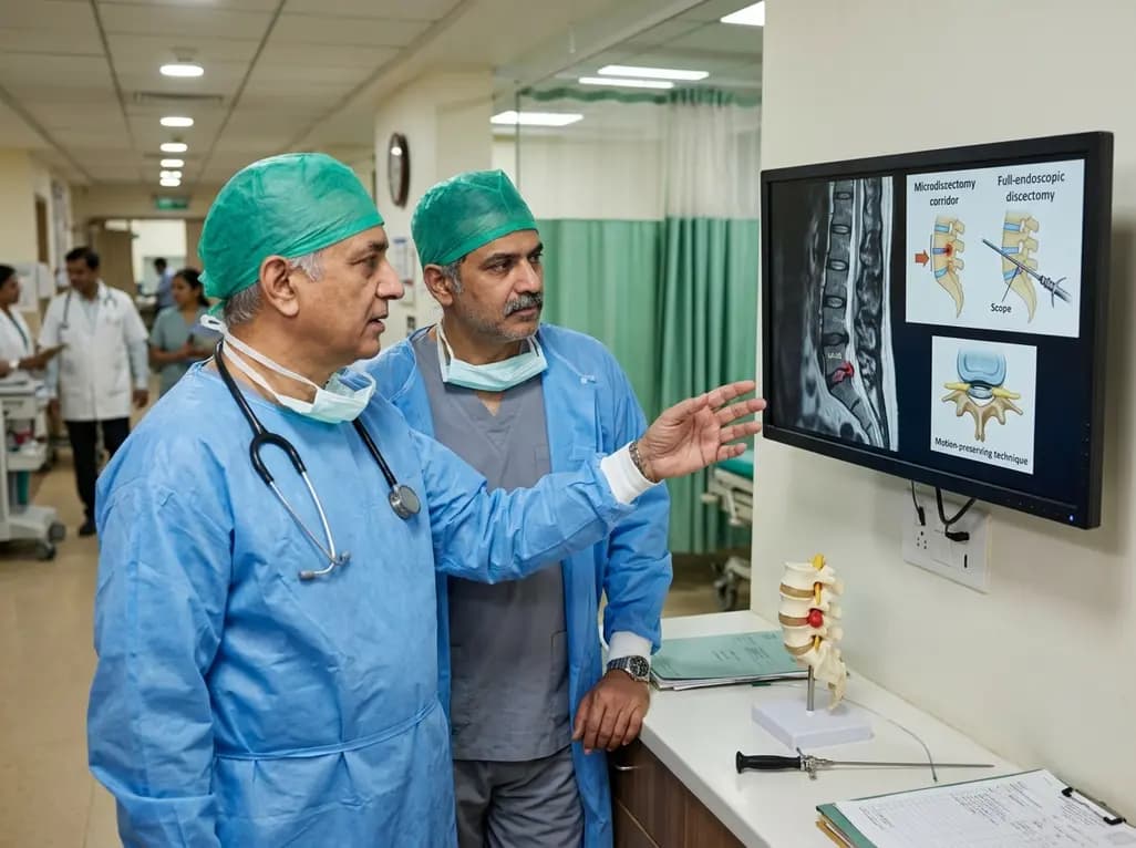
This clinical overview examines recent advancements in lumbar disc herniation surgery, comparing microscopic and full-endoscopic discectomy while highlighting biportal techniques and emerging motion-preserving innovations for spine specialists.
Today

Small interfering RNA (siRNA) therapeutics harness RNA interference to degrade disease-causing messenger RNA. By delivering hepatocyte-targeted, durable gene silencing, agents targeting PCSK9, LPA, and angiotensinogen offer potent cardiovascular risk reduction with infrequent, biannual dosing schedules.
Today
