.webp&w=384&q=75)

Loading, please wait...
Loading, please wait...

Chronic inflammatory demyelinating polyneuropathy presents substantial diagnostic challenges in everyday neurological practice. Clinicians frequently encounter progressive sensorimotor deficits that closely resemble metabolic, toxic, or hereditary neuropathies. Although electrodiagnostic testing remains the diagnostic cornerstone, atypical and purely proximal presentations often obscure definitive neurophysiological confirmation. Incomplete conduction blocks and patchy proximal demyelination regularly elude routine distal nerve conduction studies. Consequently, clinicians increasingly rely on objective supportive diagnostic modalities to avoid catastrophic treatment delays. Applying MRI in CIDP diagnosis has gained significant clinical momentum because advanced neurography directly assesses proximal spinal roots and plexuses. The blood-nerve barrier exhibits marked anatomical permeability at spinal roots and dorsal root ganglia. This unique physiological vulnerability makes these structures prime targets for early autoimmune assault. However, current international consensus guidelines historically assigned only a limited, supportive role to magnetic resonance imaging. This conservative stance stemmed from heterogeneous protocols and variable reporting standards across earlier literature. Fortunately, a recent comprehensive systematic review and meta-analysis synthesized morphological neuroimaging data across multiple international cohorts. This landmark study provides critical clarity regarding spinal root alterations, offering actionable benchmarks when electrophysiology yields equivocal results.
Qualitative evaluation on magnetic resonance neurography reveals striking structural alterations in patients with inflammatory demyelinating polyneuropathy. The systematic review identified pronounced nerve root hypertrophy, increased signal intensity on T2-weighted sequences, and post-contrast gadolinium enhancement as major hallmarks. Specifically, pooled odds ratios demonstrated that affected individuals exhibit significant nerve root thickening compared to control groups. Active inflammatory cascades trigger microvascular hyperpermeability and local endoneurial edema within spinal root sleeves. As a result, gadolinium-enhanced T1-weighted images frequently show striking contrast uptake across the cauda equina and brachial plexus. Furthermore, hyperintensity on fat-suppressed T2-weighted or short tau inversion recovery sequences highlights intense interstitial water accumulation. Repeated cycles of demyelination and aberrant remyelination drive concentric Schwann cell proliferation, forming classical histological onion bulbs. Over time, these cellular changes visibly expand individual root dimensions on cross-sectional sequences. Nevertheless, qualitative interpretation relies heavily on radiologist experience, creating potential subjective bias. Contrast enhancement may also subside rapidly after patients receive high-dose corticosteroids or intravenous immunoglobulin. Therefore, combining qualitative visual patterns with rigorous quantitative metrics ensures superior diagnostic reliability and prevents inadvertent misclassification.
Quantitative morphometry establishes objective parameters that substantially improve diagnostic specificity across diverse clinical settings. The meta-analysis analyzed extensive dimensional data and confirmed distinct anatomical thresholds for pathological enlargement. Cervical nerve root diameters averaged 5 mm in patients with CIDP, whereas lumbosacral nerve roots averaged 7 mm. Moreover, affected cohorts demonstrated significantly greater cross-sectional areas and expanded total nerve volumes than healthy controls. Interestingly, the meta-analysis demonstrated greater diagnostic significance for lumbar nerve roots compared to cervical structures. Lumbosacral roots possess a longer intradural and extraforaminal course, which provides an extended anatomical window for reliable measurement. Their larger baseline caliber also facilitates the detection of subtle pathological hypertrophy. Conversely, cervical nerve roots often suffer from physiological respiratory motion artifacts and tight bony confines within intervertebral foramina. Additionally, the standard 5 mm threshold for cervical roots overlaps somewhat with the upper limit of normal variation. In contrast, lumbosacral root enlargement exceeding 7 mm strongly discriminates inflammatory neuropathy from healthy tissue and degenerative conditions. Consequently, neurologists should prioritize lumbosacral plexus MRI when cervical spine neurography yields borderline or inconclusive findings.
Although spinal nerve root hypertrophy strongly suggests inflammatory demyelination, clinicians must carefully distinguish CIDP from various imaging mimics. Hereditary neuropathies represent the most frequent diagnostic confounder in clinical practice. Specifically, Charcot-Marie-Tooth disease type 1A produces marked, diffuse, and symmetrical nerve root hypertrophy that closely mimics chronic inflammatory neuropathy. However, hereditary neuropathies typically exhibit uniform, widespread nerve enlargement without gadolinium enhancement or clinical fluctuations. Similarly, neurofibromatosis and schwannomatosis can present with extensive nodular or plexiform nerve sheath thickening. Furthermore, advanced degenerative spine disease and lumbar canal stenosis routinely cause mechanical nerve root compression, triggering localized reactive edema. In tropical regions, chronic granulomatous infections such as neurotuberculosis or Hansen disease must remain high on the differential diagnosis. Additionally, technical variability between imaging centers introduces significant heterogeneity into quantitative measurements. Variations in slice thickness, sequence parameters, and measurement locations across neural foramina complicate comparisons. Therefore, radiologists cannot diagnose autoimmune neuropathy based solely on morphological imaging. Clinicians must always correlate neuroimaging parameters with cerebrospinal fluid protein levels, comprehensive autoantibody testing, and clinical progression before initiating immunotherapy.
Modern neuromuscular management requires a comprehensive, multi-parametric approach that combines clinical bedside acumen, electrophysiology, and advanced neuroimaging. Magnetic resonance imaging does not replace electrodiagnostic testing; rather, it functions as a powerful adjunctive modality in complex cases. Neuroimaging offers exceptional diagnostic value when patients present with atypical phenotypes, pure motor variants, or patchy proximal weakness. In these challenging presentations, routine distal conduction studies may fail to meet formal demyelinating criteria. Demonstrating nerve root hypertrophy or gadolinium enhancement satisfies supportive diagnostic criteria established by current European Academy of Neurology and Peripheral Nerve Society guidelines. Moreover, spinal MRI provides crucial reassurance when physicians contemplate long-term immunosuppression or expensive immunoglobulin maintenance therapy. Neurography also aids clinical decisions by identifying optimal sites for targeted nerve biopsies if clinicians suspect necrotizing vasculitis or neurolymphomatosis. Combining spinal MRI with high-resolution neuromuscular ultrasound creates an expansive diagnostic network that evaluates both proximal roots and distal peripheral trunks. Ultimately, prompt structural confirmation accelerates therapeutic intervention, effectively preventing irreversible axonal loss and chronic disability. Thoughtful clinical integration of high-resolution neurography directly translates into improved diagnostic accuracy and superior patient outcomes.
No, MRI cannot establish a definitive CIDP diagnosis in isolation. Magnetic resonance neurography serves as a supportive adjunctive tool rather than a standalone test. Clinicians must interpret imaging results alongside clinical signs, cerebrospinal fluid findings, and electrodiagnostic tests to exclude mimics and fulfill formal consensus diagnostic criteria.
Lumbar nerve roots offer greater diagnostic accuracy because of their larger baseline diameter and extended anatomical course. These anatomical properties facilitate clearer visualization of pathological hypertrophy. In contrast, cervical nerve roots frequently suffer from physiological respiratory motion artifacts and narrow foraminal spaces that hinder precise quantitative morphometric evaluation.
Several clinical conditions can mimic CIDP on neuroimaging, including Charcot-Marie-Tooth disease type 1A, neurofibromatosis, and neurolymphomatosis. Furthermore, spinal canal stenosis, compressive radiculopathy, and chronic infections such as neurotuberculosis cause marked root thickening. Clinicians must integrate clinical, laboratory, and electrodiagnostic features to avoid potential misinterpretation of these mimics.
Disclaimer: This content is for informational and educational purposes only and should not be taken as professional medical advice. Always consult a qualified healthcare provider for diagnosis and treatment decisions. Refer to the latest local and national guidelines for clinical practice.
References

Read summarized clinical updates, watch expert medical content, and earn CME certifications right from your smartphone.
Open your smartphone camera to scan and install.





A systematic review and meta-analysis evaluates the diagnostic utility of spinal nerve root MRI in chronic inflammatory demyelinating polyneuropathy (CIDP), demonstrating prominent hypertrophy and signal changes, particularly in the lumbosacral plexus.
Today

An Australian cohort of 34,449 adults in an unsubsidized tirzepatide digital weight loss service showed that peer-referred patients achieved significantly higher 6-month adherence (48% vs 42%) and greater weight loss (16.16% vs 14.06%) than matched non-referred peers, highlighting social support in obesity care.
Today
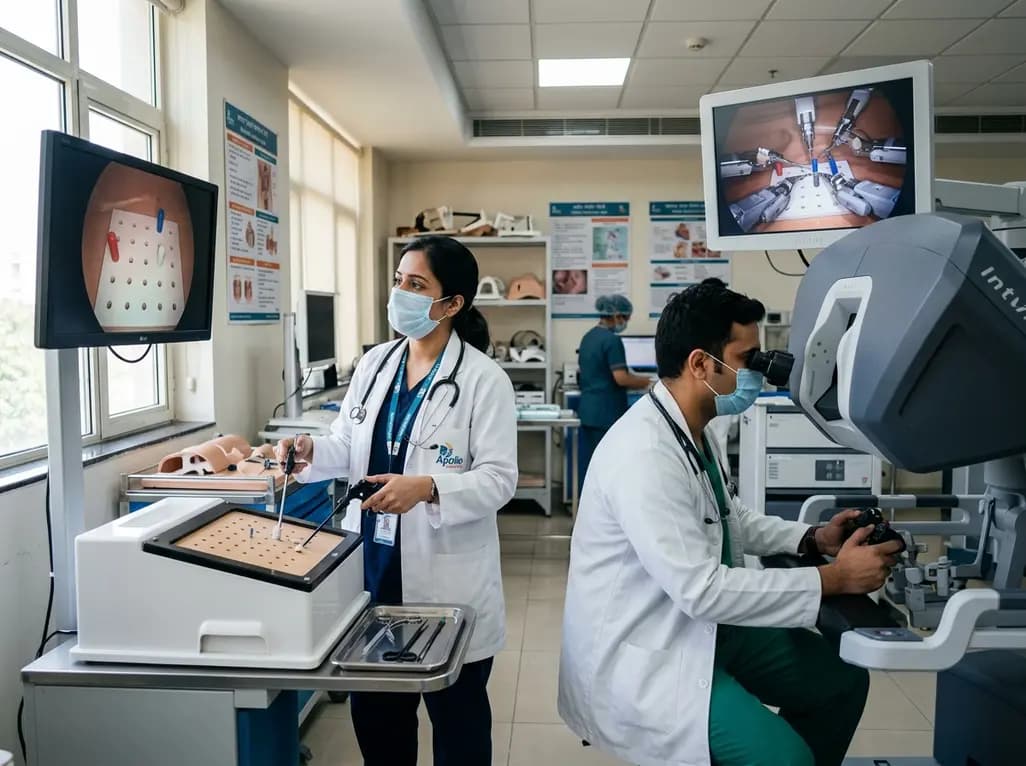
A randomized crossover study demonstrates that conventional and robotic laparoscopy require distinct neuropsychological and psychomotor skills. While trainees master conventional laparoscopy faster, robotic proficiency demands individualized, adaptive curricula tailored to specific learner profiles.
Today
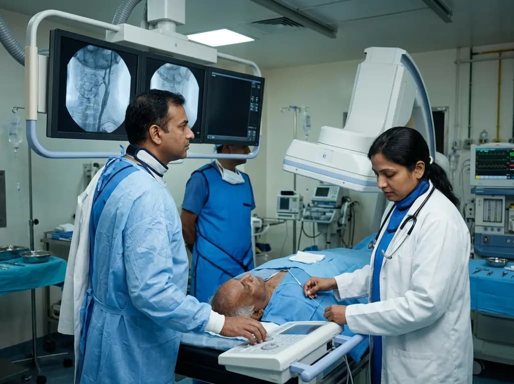
A retrospective study of 109 patients undergoing elective carotid artery stenting evaluated radiation exposure on biplane flat-panel systems. Standardized workflows and dedicated low-dose protocols provide crucial baseline data to establish neurointerventional diagnostic reference levels and optimize patient safety.
Today

A scoping review of 51 studies reveals that spinal manual therapy on the lumbo-pelvic area is commonly used for isolated lower extremity pain, predominantly knee conditions. While thrust manipulation is frequent, reporting deficits highlight the need for standardized technique descriptions to guide practice.
Today

The Supreme Court has reserved its judgment on FSSAI's proposed front-of-pack nutritional warning labels for packaged foods. With debates over compliance timelines, added versus total sugars, and ultra-processed food definitions, this landmark decision carries profound public health implications for India.
Today
