.webp&w=384&q=75)

Loading, please wait...
Loading, please wait...
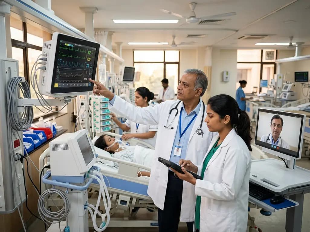
Congenital Diaphragmatic Hernia (CDH) represents a complex anatomical and physiological challenge for the neonatal intensive care team. While the primary defect is diaphragmatic, the secondary effects on pulmonary and cardiac development often dictate the clinical trajectory. Historically, management focused almost exclusively on pulmonary hypertension and lung hypoplasia. However, contemporary research increasingly highlights the critical role of neonatal CDH cardiac function in determining surgical readiness and overall survival. The study by Shalaby and colleagues provides a deep dive into the evolution of left ventricular (LV) dimensions and global cardiac performance during the perioperative period. This transition from fetal to neonatal circulation, complicated by visceral herniation, creates a unique hemodynamic environment. Consequently, understanding how the heart adapts to the sudden repositioning of abdominal contents and the subsequent changes in intrathoracic pressure is paramount. For clinicians in India, where CDH remains a significant contributor to neonatal morbidity, these insights are invaluable. By closely monitoring ventricular performance before and after repair, we can better tailor our inotropic support and fluid management strategies. This comprehensive approach ensures that the neonatal heart is not just a passive observer but an active participant in the recovery process.
In neonates with left-sided CDH, the presence of abdominal viscera in the thoracic cavity during critical stages of development leads to more than just lung compression. It often results in a smaller thoracic volume on the left side, which physically restricts the growth and filling of the left heart structures. This phenomenon, often termed relative left heart hypoplasia, is a hallmark of the disease. The compression of the left atrium and ventricle by the herniated contents reduces preload, leading to decreased stroke volume and potentially compromising systemic perfusion. Furthermore, the persistent pulmonary hypertension often seen in these infants increases right-sided pressures. This pressure shift causes a leftward deviation of the interventricular septum, which further impairs left ventricular filling and diastolic function. Understanding this interplay is essential for the medical educator and clinician alike. It is not merely a matter of anatomical displacement but a functional impairment that persists into the early neonatal period. The stabilization phase before surgical intervention is dedicated to optimizing these hemodynamics. We must balance the need for adequate systemic output with the risks of aggressive ventilation, which can further impede venous return. Recognizing the subtle signs of LV dysfunction early on can significantly improve the timing of surgical repair and the choice of perioperative support.
The surgical correction of the diaphragmatic defect is a pivotal moment that induces rapid changes in neonatal CDH cardiac function. When the herniated organs are returned to the abdominal cavity, the physical compression on the left heart is suddenly relieved. One might expect an immediate improvement in cardiac output; however, the reality is more complex. The study results indicate that while left ventricular end-diastolic and end-systolic dimensions (LVEDD and LVESD) increase significantly within 48 to 72 hours post-repair, the heart must still navigate a period of intense hemodynamic adjustment. This increase in dimensions reflects the unloading of the heart and the subsequent increase in preload as the left ventricle finally has the space to fill. Interestingly, the research shows that global cardiac performance, measured by the Tei index and systolic function parameters like MAPSE and TAPSE, improves significantly during this early post-operative window. This suggests that the heart possesses a remarkable capacity for rapid remodeling once the external mechanical constraints are removed. For surgeons and intensivists, this data underscores the importance of the first 72 hours post-surgery. It is a window of dynamic physiological flux where the cardiovascular system transitions from a state of compression to one of expansion. Monitoring these changes via echocardiography allows for real-time adjustments in vasoactive support, ensuring that the recovering heart is not overwhelmed.
The journey of a neonate with CDH does not end with the successful closure of the diaphragmatic defect. The heart continues to undergo significant remodeling long after the patient has left the operating room. The findings from the one-month follow-up are particularly illuminating. They show that the improvements in left ventricular dimensions and systolic function are not just transient post-surgical shifts but part of a sustained recovery process. Specifically, the normalization of LVEDD and LVESD z-scores suggests that the left ventricle is catching up in growth, overcoming the developmental delays imposed by the intrauterine hernia. This long-term remodeling is crucial because it ensures that the child has adequate cardiac reserve as they grow. Moreover, the study noted a concurrent improvement in right ventricular function and a reduction in pulmonary hypertension. As the pulmonary vascular resistance drops and the left heart's capacity increases, the entire cardiopulmonary unit moves toward a more stable equilibrium. This recovery highlights the importance of longitudinal follow-up. Pediatricians and cardiologists must collaborate to ensure that these infants reach their full developmental potential. Even when the initial crisis has passed, the nuances of ventricular growth and diastolic function remain relevant for several months. These insights provide a roadmap for the ongoing care of CDH survivors, focusing on the heart as a dynamic organ capable of significant post-natal adaptation.
Effective management of neonatal CDH requires a nuanced approach to vasoactive and inotropic support, guided by accurate hemodynamic monitoring. The study highlights that despite the severity of the condition, inotrope scores and vasoactive-inotrope scores (VIS) often remain stable or even decrease post-repair as cardiac performance improves. This observation is critical for clinicians who may be hesitant to scale back support in the immediate post-operative phase. By utilizing serial transthoracic echocardiography (TTE), clinicians can objectively assess changes in LV and RV function, rather than relying solely on blood pressure or heart rate. TTE provides a direct window into the heart's performance, allowing for the identification of specific issues such as septal flattening or poor ventricular contractility. In the Indian context, where resources may vary, the use of bedside ultrasound is a cost-effective and highly informative tool. It empowers the neonatal team to make data-driven decisions regarding fluid boluses, the addition of milrinone for afterload reduction, or the titration of dopamine. The goal is to facilitate the heart's natural remodeling process while maintaining adequate systemic and pulmonary perfusion. Consequently, as we refine these monitoring techniques, we can expect to see further improvements in the survival and long-term outcomes of these fragile patients. Ultimately, the heart's recovery is the engine that drives the neonate toward stability and growth.
In conclusion, the management of left-sided congenital diaphragmatic hernia has evolved from a focus on surgical technique to a comprehensive understanding of neonatal hemodynamics. The recent study by Shalaby et al. provides robust evidence that surgical repair triggers immediate and sustained improvements in neonatal CDH cardiac function and ventricular dimensions. These findings challenge the older perception that CDH is primarily a pulmonary disease. Instead, we must view it as a multisystem challenge where cardiac performance is a primary driver of success. For the Indian medical community, incorporating routine perioperative echocardiographic assessments can bridge the gap in clinical outcomes. Future research should focus on the impact of different surgical techniques on these cardiac parameters. Additionally, exploring the long-term neurodevelopmental outcomes in relation to perioperative cardiac performance would provide a more holistic view of the patient's journey. As we continue to refine our protocols, the emphasis remains on the delicate balance of pressure, volume, and space within the neonatal chest. By prioritizing the heart, we offer these infants a stronger foundation for a healthy life. Therefore, every clinical team should advocate for cardiac-focused care in CDH management to ensure the best possible start for these resilient neonates.
In neonates with left-sided Congenital Diaphragmatic Hernia (CDH), left ventricular hypoplasia primarily occurs due to the mechanical compression by abdominal organs that have herniated into the thoracic cavity. This displacement reduces the space available for the heart to develop properly during gestation. Consequently, there is limited blood flow into the left-sided chambers, which significantly impairs the growth and filling of the left ventricle, leading to reduced cardiac output and potential systemic complications post-birth.
Surgical repair of CDH alleviates the physical pressure on the heart, allowing for immediate changes in neonatal CDH cardiac function. Clinical studies show that within 48 to 72 hours post-surgery, left ventricular end-diastolic and end-systolic dimensions (LVEDD and LVESD) increase significantly. This expansion is accompanied by improvements in global cardiac performance scores and systolic function markers. These changes indicate that once the mechanical restriction is removed, the heart can rapidly adapt and initiate the necessary remodeling process.
Serial transthoracic echocardiography (TTE) is essential because it allows clinicians to monitor the heart's real-time adaptation to surgical and physiological changes. It provides critical data on ventricular filling, septal position, and global performance that standard monitoring cannot capture. By using TTE, the medical team can accurately titrate vasoactive medications and manage fluids based on the specific needs of the recovering heart. This objective assessment is vital for optimizing hemodynamic stability and improving survival rates in these complex neonatal cases.
Disclaimer: This content is for informational and educational purposes only... Refer to the latest local and national guidelines for clinical practice.
References
Shalaby MM et al. Cardiac performance and left ventricular dimensions in neonates with left-sided congenital diaphragmatic hernia before and after surgical repair. J Cardiothorac Surg. 2026 Jun 28. doi: 10.1186/s13019-026-04289-1. PMID: 42366400.
Altit G, Bhombal S, Hopper RK, et al. Management of Pulmonary Hypertension in Congenital Diaphragmatic Hernia. Pediatric Clinics of North America. 2021;68(3):651-669.
Patel N, Kipfmueller F. Cardiac Function and Hemodynamics in Congenital Diaphragmatic Hernia. Seminars in Fetal and Neonatal Medicine. 2020;25(1):101064.
"
Read summarized clinical updates, watch expert medical content, and earn CME certifications right from your smartphone.
Open your smartphone camera to scan and install.





Explore the critical changes in neonatal CDH cardiac function before and after surgical repair. This study highlights significant improvements in left ventricular dimensions and global cardiac performance within 72 hours post-surgery, offering a roadmap for better perioperative care.
3 months ago

An observational study from the NIH All of Us Research Program reveals that adding a multi-ancestry polygenic risk score to machine learning models provides only modest improvements in predicting adult weight gain, underscoring the enduring significance of routine clinical and lifestyle factors.
Today

A study in JAMIA Open shows lightweight open-source reasoning LLMs achieve up to 95% sensitivity and accelerate evidence synthesis screening by nearly twentyfold, presenting a cost-effective, scalable AI solution for biomedical research and systematic reviews.
Yesterday
A landmark study details how an enterprise pediatric remote patient management model expanded from a single cardiology pilot to 16 diverse specialty programs by combining centralized digital operations with specialty clinical leadership.
Yesterday
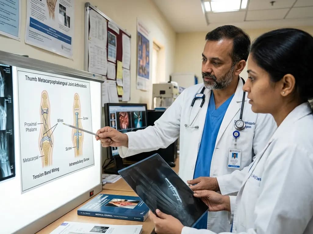
A comparative review of tension band wiring and intramedullary screw fixation in thumb metacarpophalangeal arthrodesis, evaluating union rates, hardware complications, and clinical outcomes.
Yesterday

A breakthrough ultrathin multimodal sensor decouples physical hemodynamic monitoring from disposable electrochemical calibration, achieving long-term wearable glucose prediction with 81.36% accuracy and a 9.12% MARD. This offers a robust, drift-free solution for outpatient metabolic surveillance.
Yesterday
