.webp&w=384&q=75)

Loading, please wait...
Loading, please wait...

Cerebrovascular diseases, including acute ischemic stroke and transient ischemic attacks, represent leading causes of adult disability and global mortality. Timely diagnosis remains essential for salvageable brain tissue, yet conventional emergency triage frequently experiences operational bottlenecks. Therefore, implementing multimodal stroke assessment provides an indispensable strategy to capture continuous physiological data during acute care. Traditional diagnostic protocols rely heavily on periodic physical examinations and neuroimaging suites. However, standard imaging modalities cannot provide uninterrupted real-time physiological tracking at the bedside. Wearable sensor arrays and automated artificial intelligence models directly resolve this clinical vulnerability. Furthermore, novel sensors record dynamic changes in cerebral hemodynamics, electrophysiological rhythms, and subtle motor dysfunction. Because proper early management of transient ischemic attacks prevents catastrophic completed strokes, clinicians require rapid and objective decision support. Combining non-invasive biosensors establishes a reliable quantitative baseline for acute triage teams. Consequently, physicians can identify microvascular changes before irreversible neurological deficits develop. By synthesizing electronic health records with continuous physiological measurements, hospital teams accelerate intervention intervals. Ultimately, this modern strategy improves patient survival and preserves long-term functional independence.
Modern stroke diagnostics increasingly utilize sensor fusion to examine cerebral pathophysiology through multiple complementary lenses. Specifically, researchers combine electroencephalography and near-infrared spectroscopy to monitor cortical electrical signals alongside local cerebral blood oxygenation. While electroencephalography identifies immediate neuronal slowing caused by ischemia, near-infrared spectroscopy tracks regional vascular perfusion deficits. In addition, investigators position wearable accelerometers to quantify discrete abnormalities in gait, posture, and limb movement. Automated video recording systems simultaneously capture facial asymmetry and speech patterns during standardized neurological examinations. Retinal fundus photography complements these recordings by visualizing microvascular damage that parallels intracranial cerebral microangiopathy. Thus, each sensor modality captures unique clinical information that standard physical evaluations frequently overlook. Integrating these diverse biological streams creates an exhaustive digital phenotype for every patient. Moreover, automated feature extraction algorithms interpret this multimodal stream to identify subtle functional deteriorations. Machine learning architectures then process these metrics to assist emergency clinicians with acute risk stratification. As a result, medical teams gain objective physiological measurements without subjecting vulnerable patients to invasive diagnostic procedures.
The Stroke-Data study establishes a landmark multicenter dataset to validate innovative diagnostic instrumentation across active hospital environments. Researchers conducted this prospective clinical investigation across two Finnish academic tertiary institutions: Oulu University Hospital and Kuopio University Hospital. In total, the study recruited 263 participants, comprising 103 confirmed stroke patients, 31 patients with transient ischemic attacks, and 123 healthy controls. Eligible patients enrolled within three days of symptom onset to reflect real-world acute pathophysiological dynamics. Meanwhile, healthy control participants demonstrated no history of major cerebrovascular disease, although researchers permitted maintenance antihypertensive and lipid-lowering therapies. Dedicated clinical research nurses collected comprehensive electrophysiological recordings, motion data, retinal photographs, and standardized clinical questionnaire responses. Consequently, the research team assembled a thoroughly characterized clinical cohort mirroring acute emergency presentations. Prior studies often tested individual biosensors in controlled laboratory conditions. In contrast, this multicenter protocol demonstrates practical sensor feasibility directly within inpatient stroke wards. Therefore, this public health repository provides an essential foundation for validating real-world artificial intelligence diagnostic tools.
Differentiating transient ischemic attacks from acute ischemic strokes and benign mimics represents a continuous diagnostic challenge. Transient ischemic attacks cause temporary neurological symptoms without lasting tissue infarction, meaning neuroimaging often yields completely normal findings. Nevertheless, individuals experiencing transient ischemia face an exceptionally elevated risk of sustaining debilitating strokes within subsequent days. Multimodal sensor data offer unprecedented visibility into these fleeting cerebral perturbations. For instance, continuous near-infrared spectroscopy and high-density electroencephalography detect transient perfusion deficits or localized bioelectrical slowing before conventional neuroimaging occurs. Furthermore, body-worn accelerometers document subtle kinetic asymmetries that escape routine bedside neurological examinations. Machine learning models can analyze video-recorded facial expressions and speech articulation to uncover resolving cranial neuropathies. By merging these sensitive biosignals with electronic medical records, predictive algorithms distinguish genuine vascular ischemia from non-vascular neurological mimics. Accordingly, emergency clinicians can promptly initiate secondary stroke prevention strategies, including antiplatelet regimens and vascular imaging. Consequently, precise risk evaluation during the initial seventy-two hours prevents recurring thromboembolic strokes while conserving critical inpatient resources.
Translating wearable sensor systems into standard neurological care requires streamlined deployment, minimal patient burden, and reliable data transmission. In high-pressure emergency departments, healthcare staff require intuitive instrumentation that records physiological signals within minutes. Compact wearable sensors and automated video systems fulfill this need by minimizing clinical friction while generating consistent data. Furthermore, these mobile tools extend sophisticated diagnostic capabilities into pre-hospital emergency transport and tele-stroke networks. For example, remote medical facilities lacking on-site neurologists can transmit multimodal sensor metrics directly to comprehensive stroke centers. Regional specialists then utilize automated risk scores to guide urgent thrombolysis or endovascular thrombectomy referrals. Additionally, continuous biosensing transforms outpatient stroke rehabilitation tracking. Clinicians monitor motor recovery through ambient home sensors, smart wearables, and gait metrics gathered during routine daily living. Therefore, multidisciplinary care teams adapt neurorehabilitation prescriptions based on objective functional progress rather than intermittent clinical visits. Ultimately, this connected paradigm delivers seamless longitudinal monitoring from initial emergency triage to successful community reintegration.
Traditional neuroimaging captures structural anatomical snapshots at a single point in time, which can occasionally miss transient or early-stage ischemic events. In contrast, multimodal stroke assessment combines electroencephalography, near-infrared spectroscopy, and biomechanical sensors to capture dynamic functional and hemodynamic changes continuously. This continuous physiological monitoring reveals microvascular perfusion deficits and subclinical motor abnormalities in real time. Consequently, clinical teams achieve higher diagnostic sensitivity, enabling earlier intervention before irreversible neuronal injury develops.
Transient ischemic attacks serve as an urgent harbinger of impending full-scale strokes, with up to twenty percent of patients suffering a major cerebrovascular accident shortly after presentation. Because symptoms resolve quickly, outpatient practitioners frequently underappreciate the severity of underlying vascular instability. Sensor-based data collection identifies persistent subclinical cerebral perfusion deficits and subtle motor asymmetries. Thus, rapid identification allows clinicians to initiate targeted antiplatelet therapy, carotid revascularization, and aggressive risk factor modification before permanent brain damage occurs.
Although wearable sensors demonstrate immense diagnostic potential, several technical and logistical hurdles delay universal clinical adoption. Devices require seamless interoperability with legacy electronic health record systems to avoid workflow fragmentation. Additionally, patient compliance remains variable, as frail or cognitively impaired individuals may find multiple wearable sensors uncomfortable or difficult to operate. Finally, clinicians require rigorous multicenter clinical validation demonstrating that algorithmic sensor alerts directly improve long-term functional survival without generating excessive false-positive alarms in emergency departments.
Disclaimer: This content is for informational and educational purposes only... Refer to the latest local and national guidelines for clinical practice.
References

Read summarized clinical updates, watch expert medical content, and earn CME certifications right from your smartphone.
Open your smartphone camera to scan and install.





A novel multicenter protocol establishes multimodal stroke assessment combining EEG, NIRS, motion sensors, and automated video to enhance acute triage and rehabilitation tracking.
Today
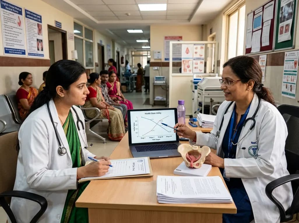
A new study evaluates health state utility for pelvic organ prolapse stages using standard gamble interviews, revealing key insights for economic modeling.
Today
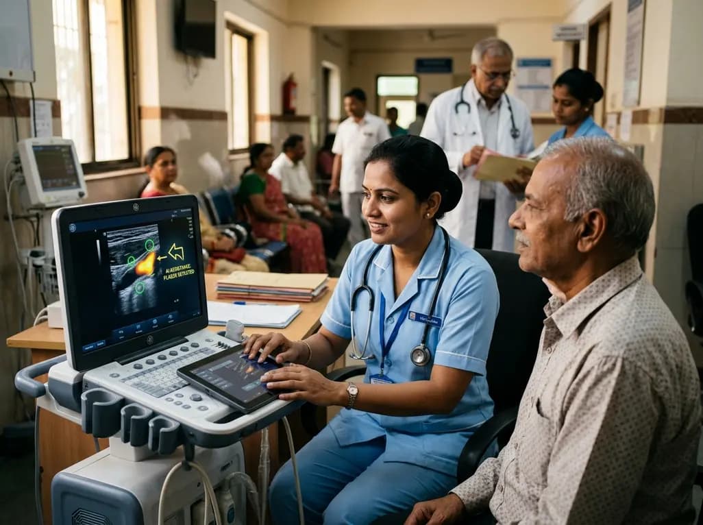
A user-centered study demonstrates that AI-assisted carotid ultrasound enables nonexpert primary care staff to detect subclinical atherosclerosis. With real-time guidance and workflow optimization, this technology supports early cardiovascular risk communication and task-shifting in routine clinical practice.
Today
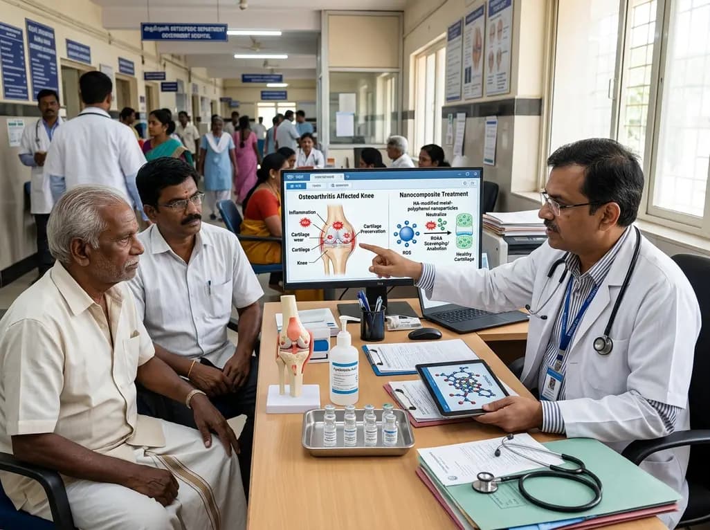
A hyaluronic acid-modified metal-polyphenol nanocomposite successfully eliminates reactive oxygen species in chondrocytes, halts cartilage breakdown, and promotes tissue anabolism, presenting a novel disease-modifying strategy for early osteoarthritis.
Today
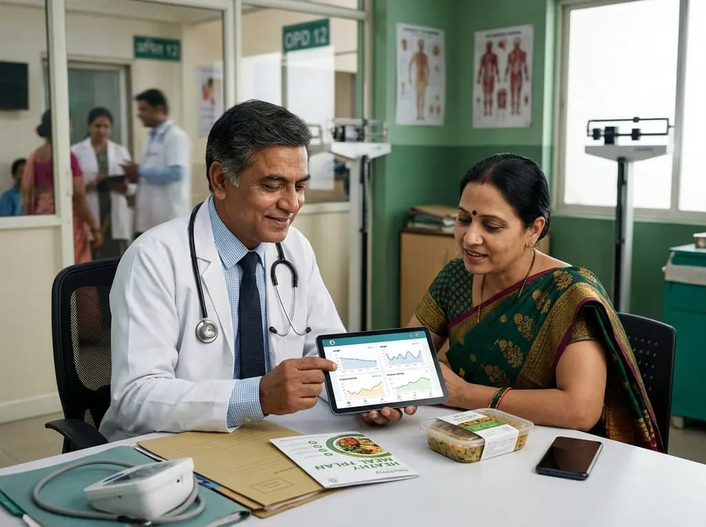
A pilot randomized trial demonstrates that digital wellness applications and medically tailored meals significantly attenuate rapid weight regain following GLP-1 receptor agonist discontinuation, providing valuable transitional support for long-term obesity management.
Today
