.webp&w=384&q=75)

Loading, please wait...
Loading, please wait...
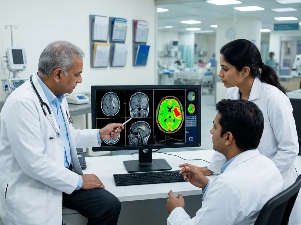
Management of glioblastoma remains one of the most significant challenges in modern neuro-oncology, particularly regarding the accurate distinction between tumor recurrence and treatment-related changes. Standard clinical practice primarily relies on Magnetic Resonance Imaging (MRI) for diagnosis and radiotherapy planning. However, MRI often faces limitations in specificity, as post-radiation effects frequently mimic active tumor growth. Consequently, the integration of multimodal PET-MR glioblastoma imaging has emerged as a vital strategy for improving diagnostic precision. Positron Emission Tomography (PET) using O-(2)-18 F-Fluoroethyl-L-Tyrosine (FET) provides unique metabolic insights that complement the structural data provided by MRI. Nevertheless, the routine adoption of this technology remains slow due to the time-intensive nature of manual segmentation and the high level of expertise required. Therefore, the development of automated models is essential to streamline clinical workflows and enhance the reliability of tumor definitions across different institutions.
Recent advancements in deep learning have introduced powerful tools for medical image analysis, with the nnU-Net framework standing at the forefront of this revolution. Researchers recently utilized this self-configuring model to automate the segmentation of tumors and organs-at-risk in a large cohort of 1,610 patients across 33 institutions. This automated approach addresses the historical bottleneck of manual contouring, which is prone to inter-observer variability. Furthermore, the model demonstrated exceptional performance, particularly in delineating the Planning Target Volume (PTV) with a Dice-Sørensen-Coefficient of 0.93. Additionally, the study successfully automated the segmentation of PET-uptake areas and MR-enhancing subregions. Resultantly, these findings suggest that AI can reliably replicate expert manual definitions, making multimodal PET-MR glioblastoma analysis more accessible for routine clinical use. Specifically, the ability to rapidly process complex imaging data allows oncologists to focus on personalized treatment strategies rather than manual data entry.
One of the most striking findings in recent neuro-imaging research is the significant spatial divergence between PET-positive areas and MRI-enhancing regions. Specifically, manual delineations of recurrence based on FET-PET and MRI often differ in both size and location. In a detailed evaluation of 185 patients, the overlap between these two modalities reached a Dice-Sørensen-Coefficient of only 0.45. This low overlap indicates that MRI and PET often capture different biological aspects of the tumor environment. For instance, FET-PET may identify metabolically active tumor cells that have not yet disrupted the blood-brain barrier enough to show enhancement on MRI. Conversely, MRI might show enhancement in areas of inflammation or radiation necrosis where active tumor cells are absent. Consequently, relying solely on a single modality may lead to incomplete tumor targeting. Thus, clinicians must integrate both structural and metabolic data to define the true extent of recurrent glioblastoma accurately.
The precision of radiotherapy planning depends heavily on the accurate definition of target volumes and the protection of surrounding healthy tissue. Automated models now provide high-fidelity segmentations for organs-at-risk (OARs), achieving a Dice-Sørensen-Coefficient of 0.70. Moreover, the integration of PET data into the planning process significantly alters the resulting treatment volumes. Since PET-positive regions often extend beyond the MR-enhancing margins, incorporating FET-PET can lead to more comprehensive coverage of the invasive tumor front. Additionally, this multimodal approach helps in sparing healthy brain tissue by identifying non-metabolic regions that may appear suspicious on MRI but do not require high-dose radiation. Resultantly, the use of automated segmentation tools ensures that these complex volumes are generated consistently, reducing the risk of geographic miss in radiotherapy. Furthermore, this standardization is crucial for multicenter clinical trials where consistency in target definition is mandatory for valid data comparison.
Radiomic feature analysis offers a sophisticated method for characterizing tumor heterogeneity beyond what is visible to the naked eye. Specifically, researchers have identified 37 distinct radiomic features that significantly differ between PET-overlapping and MRI-only enhancing regions. These features provide a deeper understanding of the tumor’s internal architecture and biological behavior. Notably, the discrimination of these features within the MR-enhancing volume showed a high sensitivity of 0.83 for predicting PET-positive subregions. This implies that even when PET imaging is not immediately available, advanced analysis of MRI radiomics may offer clues about the metabolic state of the tissue. However, the study confirms that direct PET imaging remains the gold standard for biological confirmation. By combining radiomics with automated segmentation, physicians can achieve a more nuanced view of the tumor’s invasive potential. Ultimately, this integration facilitates a more personalized approach to neuro-oncology, allowing for treatment adjustments based on the specific biological profile of the recurrence.
FET-PET provides essential metabolic information that standard MRI often lacks. While MRI identifies structural changes and blood-brain barrier disruptions, FET-PET detects increased amino acid transport, a hallmark of active tumor cells. This distinction is vital because post-treatment changes, such as radiation necrosis, often mimic tumor enhancement on MRI. By identifying metabolic hotspots, clinicians can accurately differentiate true recurrence from treatment-related alterations, ensuring patients receive appropriate salvage therapies while avoiding unnecessary, invasive interventions for non-viable tissue.
The nnU-Net framework represents a major breakthrough because it automatically configures itself to the specific characteristics of the medical imaging dataset. For glioblastoma management, this automation significantly reduces the burden on radiologists and oncologists who otherwise spend hours manually contouring tumors. Furthermore, it ensures high consistency across different clinical institutions, effectively minimizing human variability. This improved efficiency allows for faster treatment planning and more reproducible results, which are essential for both routine practice and large-scale clinical trials.
The spatial divergence, often showing an overlap of only 45%, indicates that MRI and PET capture different biological phenomena. Relying solely on MRI may lead to under-treating metabolically active areas that do not show enhancement. Conversely, it might lead to over-treating areas of pseudoprogression. Integrating both modalities allows for more precise surgical resection and more accurate radiation dosing. Consequently, this multimodal approach improves local tumor control and helps clinicians tailor therapies to the actual biological extent of the disease.
Disclaimer: This content is for informational and educational purposes only and does not constitute medical advice. Always seek the advice of a physician or other qualified health provider with any questions regarding a medical condition. Refer to the latest local and national guidelines for clinical practice.
References
Mora-Rubio A et al. Multimodal PET-MR segmentation for glioblastoma: complementarity for treatment planning and recurrence definition. Cancer Imaging. 2026 Jul 20. doi: 10.1186/s40644-026-01086-w. PMID: 42477719.
Galldiks N, Lohmann P, Albert NL, et al. Current status of PET imaging in neuro-oncology. Neurooncol Adv. 2020;2(1):vdaa071.
Isensee F, Jaeger PF, Kohl SAA, et al. nnU-Net: a self-configuring method for deep learning-based biomedical image segmentation. Nat Methods. 2021;18(2):203-211.

Read summarized clinical updates, watch expert medical content, and earn CME certifications right from your smartphone.
Open your smartphone camera to scan and install.





New research highlights how AI-driven multimodal PET-MR segmentation enhances glioblastoma management. By integrating FET-PET and MRI, clinicians can better distinguish recurrence from treatment-induced changes, leading to more precise radiotherapy planning and improved patient monitoring.
3 months ago

Discover the crucial role of glymphatic dysfunction in stroke, where perivascular clearance failure and neuroinflammation exacerbate cerebral edema and post-stroke brain injury.
Today

A prenatal case highlights severe glutaric acidemia type II presenting as bilateral hyperechoic cystic kidneys at 18 weeks. Whole-exome sequencing revealed a novel homozygous ETFDH frameshift variant, demonstrating the power of rapid genetic testing in fetal renal anomalies.
Today

Review how tailored exercise programs benefit frail older patients with heart failure. Explore pathophysiological mechanisms, individualized training prescriptions, and strategies to overcome adherence barriers in clinical practice.
Yesterday

This comparative study evaluates posterior labral tears with and without glenohumeral instability, highlighting distinct demographic patterns, MRI underestimation in stable shoulders, and successful arthroscopic repair outcomes.
Yesterday

A clinical study demonstrates that early intervention with tocilizumab, either as monotherapy or bridged with corticosteroids, achieves significant proptosis reduction and resolves active inflammation in treatment-naive thyroid eye disease.
Yesterday
