.webp&w=384&q=75)

Loading, please wait...
Loading, please wait...
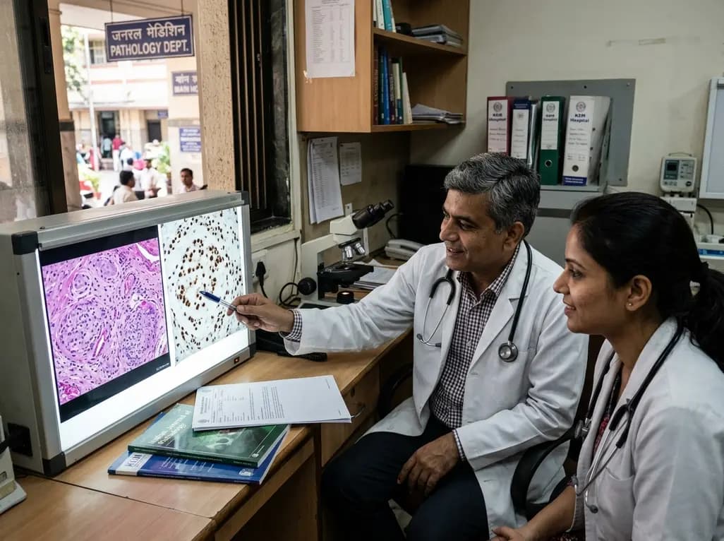
Epithelioid hemangioma represents an uncommon, benign vascular proliferation that clinicians traditionally encounter across diverse anatomical sites. Although solitary cutaneous lesions predominate in clinical practice, rare presentations exhibit multicentric clustering. Recent molecular insights demonstrate that multifocal epithelioid hemangioma frequently harbors distinct genetic alterations, particularly involving transcription factor pathways. Pathologists and dermatologists increasingly recognize that immunohistochemical FOSB overexpression clarifies diagnostic ambiguity, confirming a clonal neoplastic origin rather than an inflammatory process.
Epithelioid hemangioma can develop across various anatomical compartments, including the skin, subcutis, skeletal system, and visceral tissues. While head and neck lesions frequently affect younger individuals, elderly patients occasionally develop atypical soft tissue presentations. For instance, clinicians recently documented an elderly patient with multiple clustered lesions across the buttock and proximal thigh. Consequently, when lesions demonstrate multicentric satellitosis, clinicians often worry about malignant soft tissue sarcomas. Multifocal cutaneous lesions typically present as firm, erythematous to violaceous nodules that may occasionally ulcerate or bleed. Furthermore, deep soft tissue lesions may remain asymptomatic until they enlarge and cause localized discomfort. In addition, osseous lesions often cause localized bone pain and cortical erosion, mimicking aggressive neoplasms on cross-sectional imaging. Therefore, clinicians must maintain a high index of suspicion when evaluating multifocal vascular nodules. Diagnostic evaluation requires careful physical examination, radiological assessment, and complete tissue sampling. Because multifocal disease can simulate regional metastasis, accurate clinicopathological correlation protects patients from overtreatment or radical surgical procedures.
Microscopically, epithelioid hemangioma exhibits well-formed vascular channels lined by plump, epithelioid endothelial cells. These characteristic cells display abundant, glassy eosinophilic cytoplasm and round, vesicular nuclei with prominent nucleoli. In many instances, the endothelial cells project into the vascular lumen, creating a classic tombstone appearance. Moreover, classical lesions contain an abundant inflammatory infiltrate composed of lymphocytes, histiocytes, and eosinophils. However, cellular variants show densely packed endothelial sheets with compressed lumens and minimal inflammation. Consequently, these cellular variants often generate substantial diagnostic confusion with malignant vascular entities. Satellite nodules and multicentric growth patterns further heighten diagnostic anxiety for pathologists. Fortunately, epithelioid hemangioma lacks the destructive architectural invasion, severe pleomorphism, and atypical mitotic figures typical of angiosarcoma. In addition, pathologists rarely observe geographic coagulative necrosis in uncomplicated lesions. Nevertheless, regional multifocality and cellular exuberance mandate meticulous histological scrutiny. Pathologists must evaluate architectural boundaries, cytological details, and background stroma before establishing a final benign diagnosis.
Modern cytogenetic discoveries have fundamentally altered our understanding of vascular tumorigenesis. Historically, many pathologists considered epithelioid hemangioma a reactive inflammatory response to trauma or local vascular disruption. However, contemporary genomic sequencing reveals recurrent gene rearrangements involving the AP-1 family transcription factors. Specifically, researchers frequently identify distinct fusions involving FOS and FOSB oncogenes. Common fusion partners include ZFP36, WWTR1, and LMNA. These genetic rearrangements cause constitutive transcriptional activation, driving endothelial cell proliferation and survival. In addition, nuclear FOSB overexpression on immunohistochemistry correlates strongly with underlying chromosomal rearrangements. Interestingly, while classical variants often demonstrate FOS alterations, cellular and multifocal variants show a higher frequency of FOSB activation. Thus, immunohistochemical detection of FOSB serves as a robust surrogate marker for molecular testing. Furthermore, these recurrent alterations prove that multifocal epithelioid hemangioma represents a genuine clonal neoplasm. Understanding these molecular mechanisms enables pathologists to confirm difficult diagnoses rapidly without always requiring expensive next-generation sequencing assays.
Differentiating epithelioid hemangioma from intermediate and malignant vascular proliferations remains essential for appropriate patient management. Epithelioid hemangioendothelioma represents an intermediate-grade neoplasm characterized by cords of epithelioid cells in a myxohyaline stroma. Moreover, epithelioid hemangioendothelioma typically harbors WWTR1::CAMTA1 or YAP1::TFE3 fusions, showing nuclear CAMTA1 or TFE3 immunoreactivity. In contrast, epithelioid angiosarcoma represents a highly aggressive malignancy displaying marked cytological atypia, abundant mitoses, and infiltrative growth. Furthermore, pseudomyogenic hemangioendothelioma features spindle-to-epithelioid cells that express FOSB and keratins, but it lacks well-formed vascular lumens. Thus, an immunohistochemical panel comprising ERG, CD31, FOSB, CAMTA1, and cytokeratins facilitates an accurate differential diagnosis. In addition, Kimberly-Clark disease and cutaneous lymphoid hyperplasia enter the differential for inflammatory-rich lesions. However, these reactive conditions lack true epithelioid endothelial lining cells and genetic rearrangements. Consequently, combining detailed morphological evaluation with targeted immunohistochemistry prevents erroneous malignant diagnoses. This rigorous approach ensures that patients receive conservative management instead of aggressive chemotherapy or radiation.
The overall clinical prognosis of epithelioid hemangioma remains exceptionally favorable despite its occasional worrisome presentation. Complete surgical excision with clear margins constitutes the standard definitive therapy for accessible lesions. Because the tumor lacks metastatic potential, surgeons should avoid extensive, mutilating tissue resections. In patients presenting with multifocal or anatomically challenging lesions, clinicians may consider staged surgical excisions. Furthermore, non-surgical modalities such as pulsed dye laser, cryotherapy, and intralesional corticosteroids offer symptomatic benefit for superficial lesions. Although local recurrence occurs in approximately ten to fifteen percent of cases, re-excision typically achieves definitive cure. In addition, skeletal lesions respond reliably to curettage or marginal resection without requiring adjuvant radiotherapy. Clinicians should establish a structured post-treatment follow-up plan to monitor for local recurrence or new satellite lesions. Overall, recognizing the benign biological nature of multifocal epithelioid hemangioma prevents overtreatment. Interdisciplinary collaboration among dermatologists, oncologists, and pathologists guarantees optimal clinical outcomes and minimizes patient morbidity.
Multifocal epithelioid hemangioma displays well-formed vascular channels lined by cytologically bland, epithelioid endothelial cells with minimal mitotic activity. In contrast, epithelioid angiosarcoma exhibits marked nuclear pleomorphism, frequent atypical mitotic figures, dissecting infiltrative growth, and destructive invasion into adjacent tissues. In addition, angiosarcomas consistently lack FOSB overexpression, while epithelioid hemangiomas frequently express nuclear FOSB on routine immunohistochemical testing.
Nuclear FOSB immunostaining serves as a reliable surrogate marker for underlying FOSB gene fusions and transcript upregulation. Because malignant mimics such as epithelioid hemangioendothelioma and epithelioid angiosarcoma typically lack FOSB expression, this immunohistochemical stain clarifies challenging biopsies. Consequently, incorporating FOSB into a routine diagnostic vascular panel rapidly differentiates benign cellular epithelioid hemangiomas from aggressive malignant vascular tumors.
Complete conservative surgical excision with negative histological margins represents the primary treatment strategy for accessible multifocal lesions. Because this vascular proliferation behaves in a completely benign manner, clinicians strictly avoid radical or mutilating resections. For extensive or cosmetically sensitive cutaneous involvement, clinicians may successfully combine staged local excision with topical therapies, laser ablation, or intralesional corticosteroids to preserve tissue function.
Disclaimer: This content is for informational and educational purposes only, and does not substitute professional medical advice, diagnosis, or treatment. Clinicians must exercise independent judgment when managing vascular neoplasms. Refer to the latest local and national guidelines for clinical practice.
References

Read summarized clinical updates, watch expert medical content, and earn CME certifications right from your smartphone.
Open your smartphone camera to scan and install.





Multifocal epithelioid hemangioma is an uncommon vascular proliferation characterized by FOS/FOSB alterations, requiring precise differentiation from malignant mimics.
last month
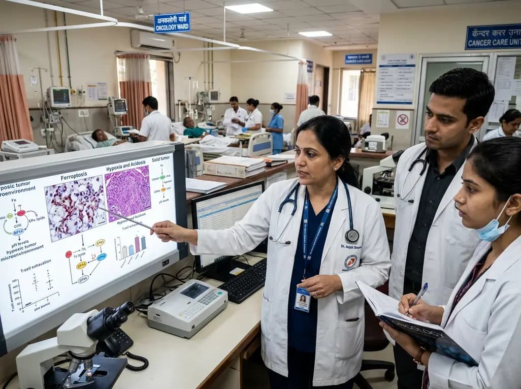
Targeting ferroptosis provides a compelling strategy to dismantle hypoxia- and acidosis-driven immunosuppression in solid tumors, enhancing response to modern immunotherapy.
Today

Findings from Project Viva indicate that sustained postpartum weight retention, rather than excessive gestational weight gain alone, directly worsens women's mid-life cardiovascular health, degrading blood pressure and glycemic parameters decades after delivery.
Today

A large UK Biobank study reveals that plasma proteomic signatures capture preclinical organ damage and improve multiorgan risk prediction across early CKM syndrome stages 0 to 2 beyond traditional PREVENT clinical models.
Today

A retrospective CBCT study reveals that superior TMJ space asymmetry correlates moderately with occipital condyle-atlas-axis asymmetry in unilateral TMJ osteoarthritis, highlighting an imaging correlate between jaw biomechanics and upper cervical alignment rather than a standalone diagnostic marker.
Today

A Bayesian multilevel meta-analysis reveals that aerobic training combined with moderate carbohydrate restriction modestly lowers HbA1c in type 2 diabetes. However, sparse data and very low certainty leave incremental benefits over exercise or diet alone unproven, highlighting the need for individualized care.
Today
