.webp&w=384&q=75)

Loading, please wait...
Loading, please wait...
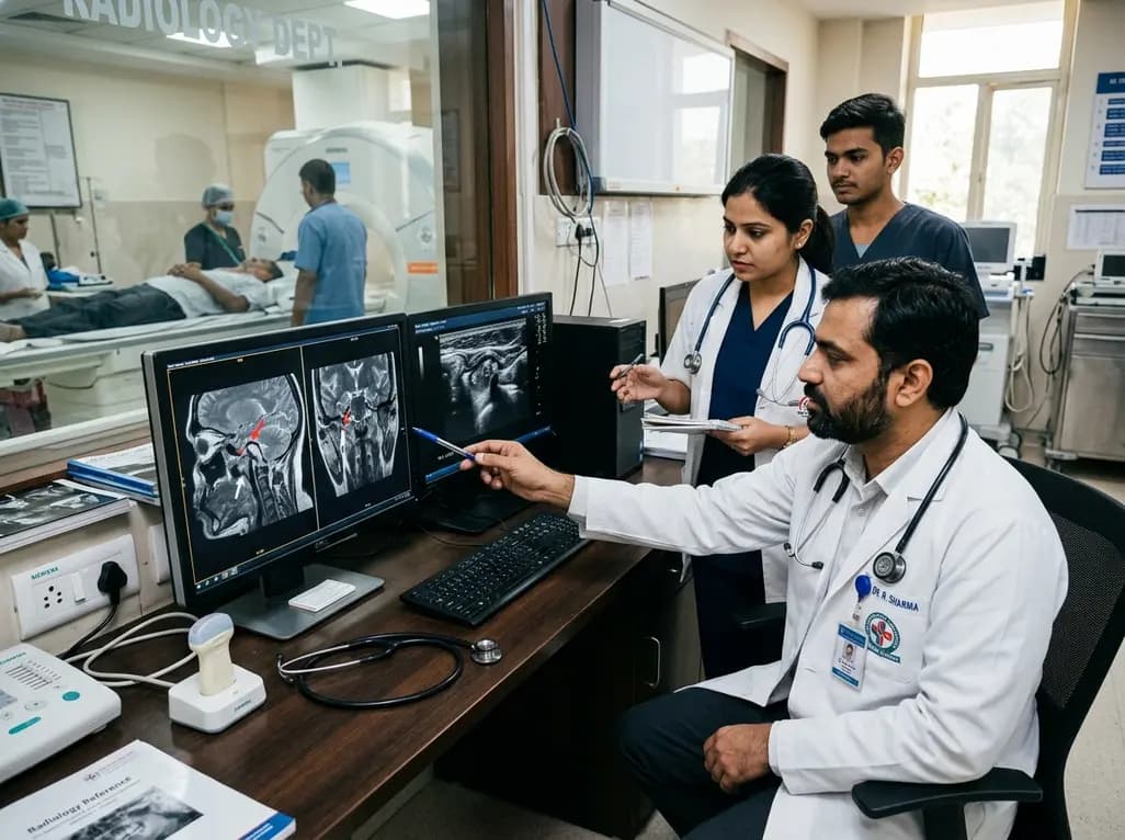
Temporomandibular disorders represent a widespread collection of musculoskeletal conditions that impair normal masticatory function and cause chronic orofacial pain. Accurate temporomandibular joint imaging serves as an indispensable clinical tool for establishing objective diagnoses and formulating targeted therapeutic regimens. Historically, clinicians relied primarily on conventional planar radiography and computed tomography to examine osseous structural integrity. However, these traditional diagnostic tools fail to visualize the articular disc, adjacent retrodiscal vascular tissue, and intra-articular fluid dynamics. Magnetic resonance imaging remains the gold standard because it delivers superior soft tissue resolution without exposing patients to ionizing radiation. Specifically, it clearly delineates the articular disc position, retrodiscal tissue integrity, cortical architecture, and intraosseous marrow alterations. Nevertheless, high examination costs, limited scanner availability, and contraindications like severe claustrophobia or metallic implants often restrict its routine clinical application in outpatient settings. In contrast, high-resolution ultrasonography has emerged as a rapid, dynamic, and non-invasive diagnostic alternative. High-frequency transducers enable real-time chairside evaluations during active mandibular opening and closing movements. Consequently, comparative studies have systematically evaluated whether sonography can match the diagnostic reliability of magnetic resonance imaging across diverse temporomandibular pathologies.
Internal derangement involving articular disc displacement constitutes the most common structural abnormality within symptomatic temporomandibular joints. Specifically, anterior disc displacement with or without reduction significantly disrupts biomechanical load distribution across the articulating surfaces. Magnetic resonance imaging accurately delineates both baseline disc position and subtle morphological deformations, such as thickening, flattening, or biconcave contour loss. Recent clinical evaluations demonstrate that high-resolution ultrasonography offers good diagnostic accuracy for detecting anterior disc displacement, particularly when patients perform active mandibular opening movements. Modern ultrasound transducers provide dynamic real-time tracking of disc translation across the articular eminence during functional jaw movements. Therefore, trained operators can easily identify disc recapturing during translation at the chairside. However, sonography exhibits clear technical limitations when evaluating rotational or sideways displacements. Medial and lateral disc displacements frequently escape sonographic detection due to acoustic shadowing from the zygomatic arch. Furthermore, subtle disc perforations, complex tears, and posterior band degenerations remain challenging to characterize completely on sonograms. Thus, while ultrasonography functions as an exceptional initial screening tool for straightforward anterior displacements, clinicians still require magnetic resonance imaging when complex multiplanar displacements or disc perforations are suspected.
Intra-articular inflammatory processes frequently manifest as joint effusion, which correlates closely with localized joint tenderness, acute arthralgia, and functional mandibular limitation. Magnetic resonance imaging detects joint effusion with remarkable diagnostic sensitivity through hyperintense fluid signals on T2-weighted sequences. Similarly, high-frequency ultrasonography reliably identifies fluid accumulation as an anechoic or hypoechoic distension within the anterior and lateral capsule compartments. Comparative diagnostic studies indicate substantial concordance between sonographic capsular widening and magnetic resonance effusion grades. In addition, chronic degenerative joint disease frequently produces progressive condylar alterations, including flattening, subchondral sclerosis, osteophyte formation, and cortical erosions. Both imaging modalities demonstrate strong diagnostic agreement when identifying prominent osteophytes and flattening along the anterior condylar surface. Nevertheless, ultrasound beams cannot penetrate dense cortical bone surfaces. Consequently, sonography cannot visualize central or posterior subchondral erosions reliably. Magnetic resonance imaging and cone-beam computed tomography provide complete three-dimensional visualization of subchondral osseous contours and structural osteoarthritic changes. Therefore, clinicians must remember that while sonography reliably detects superficial anterior osteophytes and joint effusion, comprehensive osseous staging still requires advanced volumetric or cross-sectional imaging.
Subchondral bone marrow edema within the mandibular condyle indicates acute inflammatory osteitis, elevated mechanical loading, or focal ischemic injury. On magnetic resonance imaging, marrow edema presents as diffuse hypointensity on T1-weighted sequences and prominent hyperintensity on fluid-sensitive T2-weighted fat-suppressed sequences. This pathological finding strongly correlates with acute arthralgia, rapid joint deterioration, and progressive condylar resorption. However, bone marrow edema remains completely invisible on ultrasonography because sound waves reflect entirely off intact cortical bone surfaces. Acoustic impedance prevents ultrasound beams from assessing intraosseous signal alterations or trabecular microarchitecture beneath the subchondral plate. Consequently, a patient suffering from significant condylar marrow edema may present with entirely normal osseous contours during an ultrasound evaluation. This critical diagnostic blind spot carries substantial clinical implications for overall patient management. Overlooking active bone marrow edema may lead clinicians to underestimate joint pathology or misattribute severe facial pain exclusively to muscular hyperactivity. Therefore, magnetic resonance imaging remains completely indispensable whenever clinicians suspect acute subchondral inflammation, avascular necrosis, or rapid progressive condylar resorption in symptomatic patients.
In modern dental, maxillofacial, and pain management clinics across India, establishing a tiered diagnostic workflow optimizes patient outcomes and financial resources. High-resolution ultrasonography provides substantial clinical utility as an accessible, point-of-care screening tool. Dentists and maxillofacial specialists can perform sonographic evaluations directly during outpatient consultations without lengthy delays. This chairside approach rapidly rules out gross anterior disc displacement and identifies overt joint effusion without imposing substantial financial strain on patients. Furthermore, point-of-care sonography provides real-time needle guidance for therapeutic arthrocentesis, lavage, and targeted intra-articular corticosteroid or hyaluronic acid injections with superior procedural precision. However, practitioners must clearly understand the diagnostic boundaries of sonographic assessment. When patients present with severe unremitting arthralgia, suspected bone marrow edema, or internal derangements refractory to conservative splint therapy, magnetic resonance imaging becomes mandatory. Magnetic resonance protocols provide comprehensive multiparametric visualization of soft tissue anatomy and subchondral marrow vitality. Consequently, adopting a structured diagnostic algorithm—utilizing ultrasonography for rapid screening and reserving magnetic resonance imaging for complex, refractory, or marrow-associated pathologies—ensures precise, cost-effective, and patient-centered healthcare delivery.
Ultrasonography demonstrates high diagnostic accuracy for identifying anterior disc displacement, especially during dynamic open-mouth movements. It provides rapid, real-time visualization of disc reduction at the dental chairside. However, magnetic resonance imaging remains distinctly superior for detecting rotational, medial, or lateral displacements. Furthermore, MRI characterizes subtle structural disc deformations, tear configurations, and retrodiscal tissue changes that acoustic bone shadowing prevents ultrasound transducers from evaluating effectively.
Ultrasonography cannot detect bone marrow edema because ultrasound beams reflect completely off intact cortical bone surfaces. Consequently, intraosseous pathology remains hidden. Magnetic resonance imaging remains the exclusive modality capable of visualizing condylar marrow changes. MRI reliably displays marrow edema as hypointensity on T1-weighted sequences and hyperintensity on T2-weighted fat-suppressed scans, signaling acute subchondral inflammation, mechanical overloading, or progressive condylar osteonecrosis.
Clinicians should order a dedicated MRI when patients experience severe, persistent joint pain unresponsive to conservative therapy, progressive facial asymmetry, or suspected subchondral bone marrow edema. Furthermore, MRI is essential prior to planned surgical interventions and when investigating complex multidirectional displacements or inflammatory arthritides. While sonography excels as a rapid screening tool, MRI provides comprehensive visualization of soft tissues and intraosseous vitality.
Disclaimer: This content is for informational and educational purposes only and does not constitute formal medical or dental advice. Refer to the latest local and national guidelines for clinical practice.
References

Read summarized clinical updates, watch expert medical content, and earn CME certifications right from your smartphone.
Open your smartphone camera to scan and install.





A comparative diagnostic analysis of MRI and ultrasonography for TMJ disorders, highlighting their accuracy in assessing disc displacement, effusion, condylar changes, and bone marrow edema.
last month

A recent case shows that exertional rhabdomyolysis can trigger marked transaminase spikes despite moderate creatine kinase elevation. Preserved hepatic synthetic function confirms muscle injury rather than liver failure, guiding conservative hydration and preventing unnecessary invasive diagnostics.
Today

A pharmacometric and machine learning study reveals that specific vaginal bacteria alter cervical antiretroviral exposure, highlighting the need for precision HIV PrEP strategies.
Today

Abdominal aortic aneurysm rupture remains a catastrophic vascular emergency. Groundbreaking research reveals that USP53 accelerates aneurysm progression by reprogramming smooth muscle cell metabolism toward aerobic glycolysis, highlighting a promising target beyond conventional diameter-based surveillance.
Today

A breakthrough study leverages natural language processing to accurately identify incident medication-related osteonecrosis of the jaw and track antiresorptive drug holidays from clinical narratives in electronic health records, advancing osteoporosis pharmacovigilance and dental safety.
Today

Recent research defines ferro-aging as a conserved iron-lipid axis driving organ decline across primates. Age-related iron dyshomeostasis stimulates ACSL4-mediated lipid peroxidation, promoting cellular senescence. Notably, vitamin C directly inhibits ACSL4, suppressing phospholipid oxidation and multi-organ decay.
Today
