

Loading, please wait...
Loading, please wait...

Degenerative thoracic myelopathy represents a severe yet frequently overlooked cause of progressive spinal cord dysfunction. Although clinicians routinely diagnose cervical myelopathy, degenerative thoracic lesions present distinct diagnostic and surgical hurdles. Magnetic resonance imaging remains the primary imaging modality for evaluating spinal cord compression and structural neural compromise. Recently, a systematic review published in Neurosurgical Review synthesized comprehensive imaging evidence from 59 clinical studies and 2,940 patients. This landmark analysis clarifies common radiologic patterns, underlying degenerative etiologies, and key prognostic markers. Understanding these imaging characteristics allows clinicians to detect neural compression early and optimize surgical intervention.
The systematic analysis revealed a striking anatomic distribution across the thoracic spine. Specifically, degenerative thoracic myelopathy occurred most frequently in the lower thoracic region, accounting for 62.5% of localized cases. In contrast, researchers observed a nearly equal distribution between the middle thoracic levels at 19.4% and the upper thoracic spine at 18.2%.
This predilection reflects unique regional biomechanics. The lower thoracic spine experiences heightened mobility, rotational torque, and transitional shear stress near the thoracolumbar junction. Consequently, these mechanical forces accelerate ligamentous hypertrophy and degenerative osteophyte formation. Furthermore, lower thoracic compression frequently produces overlapping neurological signs that mimic lumbar stenosis or peripheral neuropathy. Patients typically present with progressive gait ataxia, lower limb weakness, numbness, and spasticity. Therefore, physicians must maintain high vigilance when evaluating lower extremity myelopathic symptoms. Clinicians should ensure that magnetic resonance imaging includes the lower thoracic vertebrae whenever clinical findings suggest upper motor neuron dysfunction.
A central finding of the review is the clear dominance of ligamentous disorders over disc herniations. Historically, many clinicians viewed disc disease as the main driver of thoracic spinal stenosis. However, the systematic synthesis demonstrated that ossification of the ligamentum flavum represents the most prevalent etiology, affecting 71.4% of patients. In comparison, intervertebral disc herniation appeared in 20.1% of cohorts, while ossification of the posterior longitudinal ligament occurred in 19.7%.
Thus, posterior ligamentous ossification constitutes the leading structural mechanism causing thoracic spinal cord compression. Ossification of the ligamentum flavum predominantly develops along the posterolateral and dorsal canal margins, creating focal posterior dural indentation. Moreover, ligamentous ossification frequently coexists with anterior longitudinal disease, creating circumferential spinal cord compression. In addition, thoracic disc herniations often undergo secondary calcification over time, transforming soft prolapses into rigid compressive masses. As a result, neurosurgeons must utilize high-resolution magnetic resonance imaging alongside computed tomography to identify calcified components before performing decompressive surgery.
Quantitative assessment of spinal canal compromise represents an indispensable component of neuroimaging evaluations. Within the review, investigators noted that 55.5% of included studies documented objective canal measurements. Clinicians typically quantify spinal stenosis using sagittal canal diameter, transverse cross-sectional area, and canal occupancy ratios.
Importantly, the magnitude of canal compromise correlates directly with the severity of neurological impairment. When ossified ligaments and disc osteophytes occupy more than half of the spinal canal, cord functional reserve decreases rapidly. Furthermore, the thoracic canal possesses a narrow baseline diameter and vulnerable vascular supply, leaving minimal physiologic tolerance for compressive lesions. Consequently, chronic mechanical narrowing induces tissue ischemia and parenchymal damage. Clinicians also observed that circumferential stenosis causes profound neurological deficits even with moderate canal occupancy. Therefore, standardizing canal measurement protocols across radiology departments remains essential for clinical decision-making. Accurate measurements enable surgical teams to establish clear decompressive thresholds before permanent spinal cord disability occurs.
Intramedullary cord signal change serves as a critical diagnostic and prognostic biomarker in thoracic myelopathy. The systematic review identified spinal cord signal alterations on magnetic resonance imaging in 30.0% of evaluated patients. Most frequently, this manifests as focal hyperintensity on T2-weighted sequences, indicating cord edema, gliosis, demyelination, or parenchymal necrosis.
Clinically, intramedullary signal hyperintensity signals advanced microstructural distress within the spinal cord parenchyma. Patients exhibiting high T2 signal intensity present with significantly lower baseline functional scores and severe spastic gait impairment. Furthermore, the appearance of corresponding T1-weighted hypointensity denotes irreversible cavitation and permanent neuronal cell loss. In contrast, isolated faint T2 hyperintensity without T1 changes often represents reversible edema that improves following prompt decompression. Additionally, long-segment cord changes correlate with multi-level stenosis and prolonged symptom duration. Because the thoracic cord relies on a tenuous vascular network, sustained compression triggers rapid tissue ischemia. As a result, magnetic resonance imaging provides indispensable prognostic insight that guides surgical timing.
Preoperative magnetic resonance imaging provides vital indicators regarding postoperative neurological recovery. The review demonstrated that marked canal compromise, intramedullary signal hyperintensity, and multilevel involvement predict poorer functional outcomes. Similarly, ossification of the posterior longitudinal ligament portends greater surgical complexity and guarded recovery trajectories. Conversely, patients with focal single-level pathology and preserved cord signal achieve superior postoperative improvement.
Moreover, multilevel thoracic pathology complicates surgical intervention significantly. Extensive decompression increases the risk of intraoperative dural tears, cerebrospinal fluid leaks, and neurological worsening. Because ossified ligaments often adhere tightly to the underlying dura, surgeons must exercise extreme caution during decompression. Furthermore, the systematic review highlighted widespread heterogeneity in published imaging terminology and classification schemes. Consequently, the authors emphasized the urgent need for standardized radiologic criteria and uniform prospective study designs. Establishing validated imaging definitions will enhance diagnostic accuracy, streamline surgical planning, and foster collaborative global neurosurgical research.
Primary magnetic resonance imaging indicators include spinal canal compromise, dorsal compression from ligamentum flavum ossification, and ventral compression from disc herniation or longitudinal ligament ossification. Additionally, T2-weighted intramedullary cord hyperintensity occurs in nearly one-third of patients, reflecting progressive cord edema, chronic ischemia, gliosis, or irreversible myelomalacic injury.
Ossification of the ligamentum flavum predominates because the lower thoracic spine experiences substantial mechanical and rotational stress near the thoracolumbar transition zone. Furthermore, the rigid rib cage restricts upper thoracic movement, transferring biomechanical strain to lower levels. This continuous mechanical irritation drives ligamentous hypertrophy, chondrometaplasia, and progressive heterotopic ossification.
Intramedullary cord hyperintensity on T2-weighted magnetic resonance imaging correlates with more severe preoperative neurological deficits and poorer postoperative functional recovery. Furthermore, when accompanied by T1 hypointensity, it indicates irreversible cord necrosis. Consequently, patients with persistent signal changes recover less completely than individuals presenting with preserved cord signal.
Disclaimer: This content is for informational and educational purposes only and does not constitute medical advice, diagnosis, or treatment. Healthcare professionals should rely on their clinical judgment and confirmed diagnostic criteria when evaluating patients. Refer to the latest local and national guidelines for clinical practice.
References

Read summarized clinical updates, watch expert medical content, and earn CME certifications right from your smartphone.
Open your smartphone camera to scan and install.





A systematic review of 59 studies and 2940 patients identifies key MRI features in degenerative thoracic myelopathy. Ligamentous ossification predominated over disc disease, with spinal canal compromise and cord hyperintensity strongly correlating with disease severity and postoperative functional recovery.
Today

A 60-year-old woman presented with subacute cerebellar ataxia, dysautonomia, and hyperreflexia. Despite negative commercial antibody panels, tissue-based assays and phenotypic evolution to blepharospasm guided diagnostic resolution through specialized neuroimmunology testing.
Today

A prospective study reveals significant first-trimester trace-element alterations in pregnant women with epilepsy. Lower serum selenium independently predicted subsequent seizure recurrence despite prior remission, highlighting the vital clinical role of micronutrient profiling during antenatal care.
Today

A 15-year-old patient presented with knee arthralgia and acute rheumatic fever, exhibiting complex arrhythmias including AV blocks and non-sustained ventricular tachycardia despite lacking severe valvulitis. This highlights the importance of extended Holter monitoring in pediatric rheumatic carditis.
Today
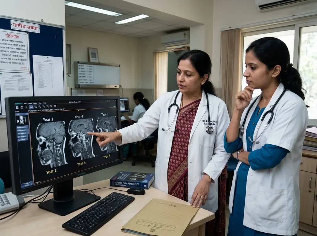
An eight-year longitudinal imaging follow-up reveals severe temporomandibular joint remodeling in nodulosis-arthropathy-osteolysis (NAO) syndrome. Despite extensive osseous destruction, the patient maintained functional jaw opening without TMJ interventions, highlighting the role of conservative management.
Today

Discover essential clinical insights into AGLP1-type amyloidosis, a rare localized complication of glucagon-like peptide-1 analog injections. Learn how histopathology and mass spectrometry differentiate benign iatrogenic amyloidomas from systemic disease, preventing misdiagnosis and unwarranted therapies.
Today
