

Loading, please wait...
Loading, please wait...

Preoperative planning remains fundamental to achieving optimal outcomes in pituitary neuroendocrine tumor surgery. Endoscopic transsphenoidal procedures provide direct access to sellar pathology, yet complex local anatomy often complicates operative execution. Surgeons routinely balance the pursuit of gross total resection against the hazardous risks of neurovascular injury and intraoperative cerebrospinal fluid leakage. Historically, surgical teams evaluated pituitary adenomas using global classification systems that often conflated lateral cavernous sinus extension with suprasellar expansion. However, contemporary skull base anatomy demonstrates that lateral and suprasellar vectors represent distinct biomechanical dimensions. Lateral invasion challenges the medial cavernous sinus wall and internal carotid artery. In contrast, superior growth stretches the diaphragma sellae and impinges upon suprasellar subarachnoid cisterns. Consequently, evaluating these anatomical vectors independently provides superior clarity for operative planning. A recent study investigated this critical paradigm using reader-concordant preoperative magnetic resonance imaging descriptors. Specifically, researchers evaluated whether lateral cavernous sinus metrics and suprasellar features show divergent associations with resection completeness versus intraoperative leakage. By separating these two axes, surgical teams can anticipate specific operative obstacles with greater precision. Therefore, comprehensive multiplanar neuroimaging evaluation provides an indispensable roadmap for operative safety.
Reliable clinical decision-making requires objective imaging markers that demonstrate minimal interobserver variability. In this multi-reader study, two blinded neurosurgeons independently reviewed preoperative magnetic resonance images from 53 consecutive patients. The evaluators assessed multiple parameters, including the modified Knosp grade, suprasellar height, bowing index, infundibular deviation angle, and a novel tumor-arachnoid cleft grade. Notably, the continuous quantitative parameters demonstrated exceptional consistency between readers. Intraclass correlation coefficients ranged from 0.995 to 0.999 for all continuous measurements. Furthermore, categorical evaluations exhibited remarkable concordant results across both independent reviewers. Exact agreement for the modified Knosp grade reached 96.2 percent, confirming its high reproducibility in experienced hands. Similarly, the study-specific tumor-arachnoid cleft grade achieved complete agreement in all 53 patients. Such outstanding concordance highlights the clinical utility of standardized radiological criteria. When multidisciplinary teams utilize unambiguous definitions, subjective measurement bias decreases substantially. Consequently, concordant magnetic resonance evaluations establish a dependable common language for skull base neurosurgeons, endocrinologists, and neuroradiologists. This high reliability is crucial because surgical risk stratification and patient counseling depend directly on these baseline imaging parameters.
Cavernous sinus involvement represents one of the most formidable hurdles to complete tumor removal. In this investigation, the modified Knosp grade emerged as the primary determinant of non-total resection. Specifically, the modified Knosp grade demonstrated an impressive area under the receiver operating characteristic curve of 0.908. After performing multivariable adjustments for maximal tumor diameter, this association remained exceptionally robust, showing an adjusted odds ratio of 10.8 per standard deviation. These quantitative results confirm that parasellar invasion into venous compartments restricts surgical resectability far more than total tumor diameter alone. However, while the modified Knosp grade strongly predicted incomplete resection, it failed to demonstrate any significant correlation with intraoperative cerebrospinal fluid leakage. The area under the curve for predicting fluid leakage was only 0.648, indicating poor discriminative performance. Therefore, surgical teams must recognize that lateral invasion dictates surgical curability but does not signal impending arachnoid disruption. In other words, cavernous sinus expansion operates independently from superior dural vulnerability. Consequently, clinicians should not use lateral invasion scores to estimate the risk of intraoperative cerebrospinal fluid leaks.
Conversely, suprasellar imaging metrics showed much stronger associations with intraoperative cerebrospinal fluid leakage, which occurred in 15 Esposito-graded events. Suprasellar height achieved an area under the curve of 0.747, while the novel tumor-arachnoid cleft grade demonstrated an area under the curve of 0.740. Remarkably, none of the 16 patients exhibiting a wide tumor-arachnoid cleft of at least 2 millimeters experienced intraoperative leakage. The presence of this visible cerebrospinal fluid buffer appears to provide a protective anatomical barrier during tumor mobilization. Statistical analyses revealed that suprasellar height and cleft grade were strongly collinear, showing a correlation coefficient of negative 0.81. Even after adjusting for maximal tumor diameter, the protective association of the tumor-arachnoid cleft persisted with an odds ratio of 0.43. Hence, superior expansion that obliterates the arachnoid cleft markedly elevates intraoperative breach risk. Although these findings remain exploratory, they explain why certain large tumors separate smoothly from the optic chiasm without causing fluid leakage. Furthermore, identifying this fluid layer helps neurosurgeons anticipate which tumors have breached the sellar diaphragm.
Integrating these dual anatomical axes directly optimizes operative tactics and patient counseling. When preoperative imaging displays high modified Knosp grades, neurosurgeons should anticipate residual tumor tissue. In functional adenomas, the multidisciplinary team can plan adjuvant stereotactic radiosurgery or targeted medical therapy well in advance. Moreover, surgeons can utilize angled endoscopes and micro-doppler probes to safely inspect the cavernous sinus medial wall. Conversely, when imaging reveals prominent suprasellar height accompanied by cleft obliteration, surgeons must anticipate high-flow cerebrospinal fluid leaks. Under these conditions, the surgeon must avoid aggressive downward traction on the tumor capsule, which could tear an attenuated diaphragma. In addition, the operative team should proactively prepare multilayered skull base reconstruction strategies before initiating tumor debulking. Using autologous fascia, fat grafts, and vascularized nasoseptal flaps provides durable cranial base repair, thereby preventing postoperative pseudomeningoceles and meningitis. Although the tumor-arachnoid cleft metric requires prospective external validation in multicenter cohorts, this dual-axis evaluation framework substantially refines surgical risk stratification. Ultimately, separating lateral resectability from superior leakage risks elevates surgical safety and patient outcomes.
The tumor-arachnoid cleft represents a visible cerebrospinal fluid buffer separating the superior tumor margin from the suprasellar arachnoid. A wide cleft measuring at least 2 millimeters indicates preserved arachnoid integrity, which significantly reduces the risk of intraoperative cerebrospinal fluid leakage during endoscopic transsphenoidal tumor mobilization and resection.
The modified Knosp grade evaluates lateral cavernous sinus extension and carotid artery relationships rather than superior diaphragma sellae integrity. Because lateral parasellar expansion does not correlate with thinning or tension across the suprasellar arachnoid membrane, high Knosp grades predict incomplete tumor resection without reliably forecasting intraoperative cerebrospinal fluid leakage.
Neurosurgical teams utilize suprasellar height to anticipate diaphragma sellae attenuation and superior cistern herniation. Elevated suprasellar extension frequently correlates with cleft obliteration and higher tear vulnerability. Consequently, identifying pronounced suprasellar height prompts preemptive preparation of multilayered skull base reconstruction materials, such as vascularized nasoseptal flaps and autologous tissue grafts.
Disclaimer: This content is for informational and educational purposes only... Refer to the latest local and national guidelines for clinical practice.
References

Read summarized clinical updates, watch expert medical content, and earn CME certifications right from your smartphone.
Open your smartphone camera to scan and install.





Preoperative MRI evaluation of lateral Knosp grade and suprasellar tumor-arachnoid anatomy predicts distinct outcomes in pituitary neuroendocrine tumor surgery. While Knosp grade strongly dictates resection completeness, suprasellar height and cleft grade correlate with intraoperative CSF leak risk.
Today

Gastrointestinal smooth muscle cells rely on nanoscale membrane domains to integrate mechanosensitive currents. Recent insights show that nanodomain architecture, rather than channel properties alone, orchestrates motility, opening new therapeutic avenues for clinical dysmotility and gastrointestinal aging.
Today

A landmark study reveals that lipid-rich malignant ascites alters the T-cell metabolic transcriptome, causing cholesterol efflux and impaired fatty acid oxidation. This suppresses TCR signalling and blocks BiTE immunotherapy, while lipid depletion restores antitumour cytotoxicity in ovarian cancer.
Today
Novel clinical evidence demonstrates the feasibility of combining VA-ECMO with the NyokAssist self-expanding transvalvular pump for active left ventricular unloading in infarct-related cardiogenic shock, achieving successful stabilization and decannulation.
Today
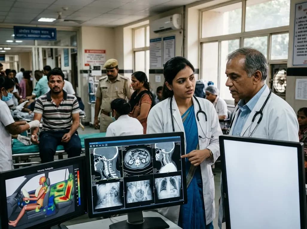
Recent crash analyses demonstrate significant variations in traffic injury severity across vehicle seating positions. Electric vehicle passengers face disproportionately higher trauma risks than drivers, particularly during airbag deployments, demanding adapted emergency assessment and clinical vigilance.
Today

Understand the causal role of Lipoprotein(a) in cardiovascular disease, why international guidelines recommend once-in-a-lifetime molar testing, and the clinical implications of recent phase 3 RNA outcome trials.
Today
