.webp&w=384&q=75)

Loading, please wait...
Loading, please wait...
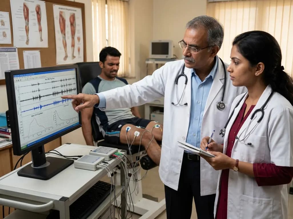
Neuromuscular fatigue represents a complex physiological phenomenon involving both central drive and peripheral contractile failure. During maximal isometric contractions, the central nervous system must continuously modulate motor unit firing rates to sustain force production. Clinicians and exercise physiologists have long debated whether biological sex influences the rate of neural drive degradation during exhaustive exertion. A seminal neurophysiological investigation examined maximal motor unit firing rates within the quadriceps femoris to clarify these responses. Researchers recorded intramuscular single motor unit activity from the deep vastus intermedius and superficial vastus lateralis muscles in healthy males and females. The experimental protocol utilized a sustained one-minute maximal voluntary isometric contraction followed by a structured five-minute recovery timeline. Throughout this demanding task, voluntary force and neural discharge frequencies experienced a profound decline of approximately fifty percent. Interestingly, despite known morphological differences in skeletal muscle architecture between sexes, the relative drop in neural drive remained uniform across both cohorts. Therefore, these electrophysiological data demonstrate that motor unit discharge modulation during severe fatigue follows a highly conserved physiological pattern.
The quadriceps femoris acts as the primary extensor mechanism of the human knee joint, playing a vital role in locomotion, athletic performance, and postural balance. When an individual sustains a maximal voluntary contraction, metabolic byproducts rapidly accumulate within active muscle fibers. Consequently, inorganic phosphate and hydrogen ions impair cross-bridge kinetics and reduce sarcoplasmic calcium sensitivity. Simultaneously, afferent feedback from group III and IV sensory fibers increases, sending inhibitory signals to spinal motor neurons and supraspinal motor centers. This regulatory feedback loop leads to a progressive reduction in motor unit firing rates, preventing irreversible cellular damage from catastrophic energetic depletion. Electrically evoked doublet stimulation at one hundred hertz confirmed substantial peripheral contractile impairment alongside central discharge slowing. However, the degree of force depression mirrored the drop in neural firing rates across all participants. In addition, the parallel reduction across deep and superficial quadriceps compartments highlights a unified neuromuscular fatigue threshold. Neurologists and physical medicine practitioners must recognize that central discharge slowing functions as a protective muscular strategy rather than isolated neuromuscular failure.
Historically, physiological studies suggested that females exhibit greater muscular endurance during submaximal isometric tasks due to a higher proportion of type I muscle fibers and greater capillary density. However, this endurance advantage often diminishes during sustained all-out maximal voluntary contractions. In this investigation, both male and female participants demonstrated nearly identical proportional decreases in motor unit firing rates over the sixty-second maximal knee extension. Furthermore, during the subsequent five-minute rest period, both sexes exhibited a rapid and equivalent recovery trajectory of neural discharge frequencies. Contractile force and evoked twitch parameters also recovered at comparable rates across genders. Therefore, while sex differences in absolute strength and muscle cross-sectional area remain evident, the intrinsic central motor drive dynamics appear sex-neutral under maximal isometric strain. Understanding these uniform neural characteristics helps clinicians design equitable training regimens and evidence-based rehabilitation protocols. By acknowledging that central fatigue mechanisms operate similarly in men and women during maximal exertion, sports medicine specialists can optimize physical therapy regimens without presuming disparate neural fatigue vulnerabilities.
Evaluating intramuscular electromyography from multiple synergist muscles provides vital information regarding spatial recruitment strategies during exhaustive efforts. The vastus intermedius lies deep beneath the rectus femoris and vastus lateralis, serving as a primary monoarticular knee extensor with substantial postural responsibilities. In contrast, the vastus lateralis frequently generates higher peak forces during dynamic lower limb tasks. Despite their divergent anatomical positions and fiber orientations, both muscles showed remarkably synchronized declines in motor unit firing rates during sustained maximal contraction. High-resolution ultrasound guidance ensured precise intramuscular tungsten microelectrode placement, isolating discrete single-unit action potentials without signal crosstalk. Consequently, the findings indicate that central descending drive distributes neural excitation equitably across both deep and superficial synergists during maximal demand. Furthermore, the recovery kinetics of single motor units in both muscles aligned closely throughout the five-minute post-contraction monitoring phase. Thus, neuromuscular rehabilitation targeting quadriceps strengthening can rely on synergistic neural adaptations across the entire muscle group rather than assuming localized neural sparing.
These neurophysiological insights hold significant practical relevance for clinicians managing athletic conditioning, postoperative knee rehabilitation, and neurological disorders. Because maximal motor unit firing rates decline swiftly within sixty seconds, therapeutic protocols should carefully structure contraction durations to avoid excessive neural exhaustion. High-intensity resistance training designed to stimulate neuromuscular adaptations requires adequate inter-set rest intervals of at least three to five minutes. During this timeframe, central motor discharge frequencies and twitch potentiation recover substantially toward baseline levels. Moreover, orthopedic surgeons and physiotherapists rehabilitating patients after anterior cruciate ligament reconstruction or total knee arthroplasty can apply these principles directly. Since male and female neuromuscular systems exhibit equivalent recovery timelines following maximal output, clinicians do not need sex-segregated rest intervals for maximal isometric conditioning. Instead, practitioners should focus on progressive overload, proper biomechanical alignment, and targeted muscle activation. In addition, monitoring rate of force development and voluntary activation helps identify subtle central drive deficits in recovering athletes.
Implementing evidence-based recovery strategies is essential for maximizing athletic performance and minimizing overuse injuries. Post-exercise recovery encompasses both peripheral metabolic clearance and the restoration of central descending motor drive. Because motor unit discharge rates rebound significantly within several minutes post-contraction, structured interval training can maintain high motor recruitment while avoiding chronic neuromuscular burnout. Furthermore, sports scientists should incorporate active recovery techniques, appropriate hydration, and adequate carbohydrate replenishment to support peripheral muscular restoration alongside central neural recovery. Clinicians working with high-performance athletes must also account for cumulative training fatigue, which can impair neural drive over multi-day training cycles. By evaluating electrophysiological properties alongside subjective fatigue scores, strength and conditioning coaches can fine-tune training volumes with greater precision. Ultimately, recognizing that maximal motor unit firing rates follow predictable degradation and recovery pathways empowers clinicians to enhance muscular performance across diverse patient populations safely and effectively.
Current electrophysiological evidence demonstrates that men and women experience comparable declines in motor unit firing rates during sustained maximal isometric contractions. Although absolute force production and muscle cross-sectional area vary between sexes, the relative reduction and subsequent recovery of central neural discharge remain virtually identical. Consequently, clinicians do not need to prescribe disparate rest intervals based solely on biological sex during high-intensity maximal isometric training programs.
Motor unit discharge frequencies decline due to a combination of central and peripheral regulatory mechanisms. As metabolic byproducts accumulate in contracting muscle fibers, inhibitory group III and IV sensory afferents send feedback to spinal and supraspinal motor circuits. Consequently, this protective neural reflex slows central motor drive, reducing motor unit firing rates to prevent severe cellular energetic crises and structural damage within the working skeletal muscle tissue.
Maximal motor unit firing rates exhibit a rapid initial recovery within the first two to three minutes following exhaustive isometric exertion, approaching near-baseline values by five minutes. However, full recovery of maximal voluntary force may take longer due to lingering peripheral contractile fatigue. Therefore, incorporating structured rest intervals of three to five minutes between maximal sets ensures optimal neural drive restoration in neurorehabilitation protocols.
Disclaimer: This content is for informational and educational purposes only. It is not intended to be a substitute for professional medical advice, diagnosis, or treatment. Always seek the advice of your physician or other qualified health provider with any questions you may have regarding a medical condition. Refer to the latest local and national guidelines for clinical practice.
References

Read summarized clinical updates, watch expert medical content, and earn CME certifications right from your smartphone.
Open your smartphone camera to scan and install.





A study evaluating motor unit firing rates in the vastus intermedius and lateralis during maximal knee extensions revealed comparable fatigue-induced declines and recovery trajectories across sexes, refining clinical perspectives on neuromuscular fatigue and rehabilitation.
last month

With post-monsoon dengue surges threatening South Asian health systems, physicians must refine triage and management. This guide covers vital warning signs, plasma leakage detection, crystalloid titration, and vector containment strategies to prevent mortality during seasonal arboviral epidemics.
Today

Findings from Project Viva indicate that sustained postpartum weight retention, rather than excessive gestational weight gain alone, directly worsens women's mid-life cardiovascular health, degrading blood pressure and glycemic parameters decades after delivery.
Today

A large UK Biobank study reveals that plasma proteomic signatures capture preclinical organ damage and improve multiorgan risk prediction across early CKM syndrome stages 0 to 2 beyond traditional PREVENT clinical models.
Today

A retrospective CBCT study reveals that superior TMJ space asymmetry correlates moderately with occipital condyle-atlas-axis asymmetry in unilateral TMJ osteoarthritis, highlighting an imaging correlate between jaw biomechanics and upper cervical alignment rather than a standalone diagnostic marker.
Today

A Bayesian multilevel meta-analysis reveals that aerobic training combined with moderate carbohydrate restriction modestly lowers HbA1c in type 2 diabetes. However, sparse data and very low certainty leave incremental benefits over exercise or diet alone unproven, highlighting the need for individualized care.
Today
