

Loading, please wait...
Loading, please wait...

Orthopaedic trauma surgeons frequently encounter complex rotational injuries of the distal tibia and fibula. Among these patterns, PER ankle fractures present substantial diagnostic challenges, particularly regarding occult intra-articular extensions. While clinicians traditionally rely on standard three-view radiographs, conventional plain films often conceal subtle articular impaction. Consequently, surgeons may misjudge the true anatomical complexity of the distal tibial articular surface. A landmark multi-institutional investigation by Lee and colleagues critically evaluated how accurately conventional radiographs detect anterolateral tibial plafond involvement compared with computed tomography. Their compelling findings highlight critical diagnostic blind spots that every practicing orthopaedic specialist must address to prevent debilitating post-traumatic ankle arthrosis.
Pronation-external rotation injuries, classified under the AO/OTA 44C schema, classically result from high-energy rotational mechanisms. In these severe injuries, torsional forces traverse sequentially through the anterior syndesmotic complex, the fibular shaft, and the posterior structures. However, trauma specialists increasingly recognize that energy also propagates across the weight-bearing tibial plafond. Specifically, anterolateral articular rim fracture lines or direct chondral impactions frequently develop alongside classic syndesmotic ruptures. In many clinical presentations, these lesions mimic the well-known Tillaux-Chaput fragment. Yet, they often involve depressed articular cartilage that requires anatomical elevation. If clinicians miss this articular depression during initial preoperative assessments, incongruence inevitably persists within the ankle mortise. As a result, unaddressed joint step-offs accelerate uneven peak contact pressures across the talar dome during weight-bearing activities. Furthermore, unrecognized intra-articular step-offs predispose active individuals to early-onset osteoarthritis, persistent instability, and chronic debilitating pain. Therefore, orthopaedic trauma teams must maintain a heightened suspicion for anterolateral articular disruption whenever assessing high-energy rotational malleolar disruption. Identifying these subtle articular fractures early prevents long-term joint destruction.
To resolve longstanding diagnostic uncertainties, researchers conducted a comprehensive multi-institutional retrospective review across two level 1 trauma centers in the United States. The investigators evaluated eighty-three adult patients who underwent operative treatment for PER ankle fractures between 2020 and 2022. Every patient had adequate preoperative standard radiographs, comprising anteroposterior, mortise, and lateral ankle projections. In addition, a paired subset of thirty-seven patients underwent both standard plain radiography and advanced axial computed tomography. Four fellowship-trained orthopaedic trauma surgeons independently scrutinized each radiographic series to determine fracture involvement and true impaction. Crucially, researchers established computed tomography as the benchmark gold standard to judge plain film diagnostic utility. Moreover, the investigators tracked whether reviewing subsequent cross-sectional scans directly altered the surgeons' stated intent to execute open reduction and articular visualization. By adopting this rigorous multi-reviewer methodology, the authors systematically uncovered the hidden threshold where standard radiography fails to depict articular topography accurately. Their objective assessment exposed crucial discrepancies between conventional planar imaging and high-resolution volumetric scans.
The study results revealed striking diagnostic limitations inherent to standard plain radiography. Among the entire cohort of eighty-three patients, initial plain radiographs detected anterolateral tibial plafond involvement in only 14.5% of cases. Similarly, plain films identified articular impaction in merely 4.8% of these cases. However, when analyzing the thirty-seven patients with paired advanced imaging, computed tomography revealed true plafond involvement in 37.8% of cases. Plain radiographs only detected involvement in 13.5% of this group, demonstrating a poor sensitivity of just 21%. Furthermore, computed tomography verified true articular impaction in 8.1% of patients, whereas plain radiographs detected impaction in only 5.4%. Consequently, radiographs achieved a modest sensitivity of 67% for impaction, although their specificity reached an impressive 100%. While plain films exhibited high positive predictive values, their negative predictive values failed to provide clinical security. In addition, the interobserver reliability among fellowship-trained trauma surgeons assessing plain radiographs remained notably poor. Therefore, relying exclusively on plain radiographs risks leaving significant joint depression unrecognized. Orthopaedic surgeons frequently fail to spot articular fragmentation on standard radiographic views.
Accurate identification of anterolateral plafond depression dictates critical shifts in operative tactics. In this investigation, plain radiographs predicted surgeons' stated intent to pursue direct visualization and articular reduction in only 66.7% of cases. Consequently, one in three patients requiring direct joint restoration would have received inadequate exposure without cross-sectional imaging. When surgeons identify depressed anterolateral fragments, they typically must elevate and bone-graft the impacted subchondral bed. Moreover, obtaining rigid internal fixation requires distinct anterior or anterolateral surgical exposures rather than standard lateral approaches. Without cross-sectional imaging, surgeons frequently rely on indirect reduction methods through lateral fibular incisions. However, indirect syndesmotic clamping cannot reliably reduce depressed intra-articular plafond fragments. Thus, missing these impacted osteochondral fragments leads to persistent syndesmotic malreduction and articular incongruity. By introducing computed tomography early in the workup, surgical teams formulate definitive approaches that optimize direct articular visualization. Preoperative computed tomography fundamentally transforms surgical planning from presumptive maneuvers into precise, patient-specific interventions.
These compelling radiographic findings have immediate clinical implications for high-volume emergency departments and orthopaedic centers. Because severe rotational ankle injuries often occur during motor vehicle accidents and athletic activities, clinicians must recognize high-risk patterns promptly. Standard radiographic series remain invaluable for initial alignment assessments and gross fracture triage. Nevertheless, plain films clearly lack sufficient sensitivity to exclude complex anterolateral articular injury. Although obtaining computed tomography for every isolated ankle fracture may strain health resources, high-energy rotational patterns clearly warrant advanced imaging. Specifically, whenever clinicians observe significant fibular displacement, severe syndesmotic diastasis, or complex posterior malleolar involvement, they should maintain a low threshold for advanced scans. Furthermore, obtaining timely cross-sectional imaging avoids unexpected intraoperative findings that can compromise operating room efficiency. Surgeons who utilize cross-sectional imaging can tailor patient counseling and surgical timing more effectively. Ultimately, integrating advanced imaging into diagnostic protocols ensures that orthopaedic surgeons achieve true anatomical restoration and superior long-term patient outcomes.
Anterolateral tibial plafond involvement refers to any discrete fracture line or articular impaction disrupting the anterolateral distal tibial joint surface. In pronation-external rotation mechanisms, traumatic forces propagate across the anterior syndesmosis, fracturing or depressing the subchondral bone. While avulsions resemble Chaput fragments, true impaction involves crushed articular cartilage. Therefore, surgeons must distinguish between simple ligamentous avulsions and true articular depressions that require surgical elevation and stable subchondral bone grafting.
Standard plain radiographs frequently miss anterolateral impaction because overlapping osseous structures, such as the distal fibula and tibial margins, obscure subtle articular steps. Furthermore, two-dimensional projections cannot adequately capture complex three-dimensional rotational topography. The study showed plain radiography achieved an unacceptable sensitivity of only 21% for involvement. Consequently, clinicians who rely solely on plain films frequently overlook depressed osteochondral fragments, which compromise joint congruency and accelerate degenerative post-traumatic arthritis.
Surgeons should routinely obtain preoperative computed tomography whenever they evaluate high-energy rotational ankle injuries, including pronation-external rotation fractures. Furthermore, scans are essential when radiographs reveal posterior malleolar fragments, syndesmotic widening, or questionable articular impaction. Because plain radiographs demonstrate poor interobserver reliability and low diagnostic sensitivity, advanced cross-sectional imaging provides critical anatomical clarity. Consequently, preoperative computed tomography directly guides appropriate surgical exposure, anatomical joint reduction, and definitive subchondral stabilization.
Disclaimer: This content is for informational and educational purposes only and does not constitute medical advice, diagnosis, or treatment. Refer to the latest local and national guidelines for clinical practice.
References

Read summarized clinical updates, watch expert medical content, and earn CME certifications right from your smartphone.
Open your smartphone camera to scan and install.





A multi-center study reveals that plain radiographs demonstrate poor sensitivity for detecting anterolateral tibial plafond involvement in pronation-external rotation (PER) ankle fractures. Routine preoperative CT imaging ensures accurate identification of articular impaction and guides surgical reduction.
Today
Understand the causal role of Lipoprotein(a) in cardiovascular disease, why international guidelines recommend once-in-a-lifetime molar testing, and the clinical implications of recent phase 3 RNA outcome trials.
Today
Perimenopause brings erratic hormonal surges and metabolic shifts that significantly affect breast tissue. This comprehensive clinical review examines benign breast conditions, cancer risk stratification, mammographic density challenges, and endocrine therapy transitions in midlife women.
Today
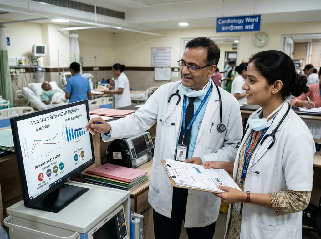
The VOCAL-AHF study reveals that most acute heart failure patients across reduced and non-reduced ejection fraction categories are eligible for in-hospital GDMT initiation. Notably, nearly two-thirds of HFrEF patients qualify for quadruple therapy, while non-reduced EF patients show high eligibility for dual therapy.
Today

This review examines the pharmacogenomics of antidiabetic agents, highlighting how genetic variations in SLC2A2, SLC47A1, CYP2C9, ABCC8, and GLP1R influence drug transport, therapeutic efficacy, and hypoglycemia risk to enable tailored precision medicine in type 2 diabetes.
Today
