.webp&w=384&q=75)

Loading, please wait...
Loading, please wait...
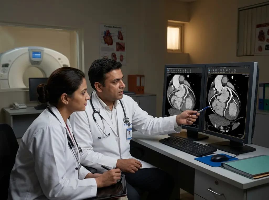
Coronary computed tomography angiography (CCTA) serves as an essential non-invasive imaging modality for evaluating coronary artery disease across diverse patient populations. However, rapid cardiac motion and elevated heart rates frequently create troublesome blurring and stair-step artifacts. These artifacts compromise image interpretability, especially when the mid-diastolic rest period becomes abbreviated. Applying an advanced motion-correction algorithm offers an innovative computational solution to overcome these diagnostic obstacles. Traditionally, clinicians rely heavily on pharmacological heart rate control using oral or intravenous beta-blockers to prolong diastolic quiescence. Unfortunately, numerous patients present with clinical contraindications, such as severe asthma, acute bronchospasm, or severe hypotension. Furthermore, some individuals fail to achieve target heart rates despite premedication. Advanced reconstruction algorithms analyze raw projection data to counteract coronary displacement dynamically. Consequently, this technology enhances image clarity without altering patient cardiac physiology or requiring extra scan acquisitions. By recovering diagnostic evaluability in motion-affected scans, healthcare teams avoid unnecessary repeat examinations and excessive radiation exposure. Therefore, implementing motion correction directly addresses a fundamental challenge in everyday cardiovascular radiology and improves overall workflow efficiency.
Modern 320-detector row CT scanners provide complete anatomical volume coverage of the heart within a single gantry rotation. Even with rapid rotation speeds of 0.24 seconds, residual cardiac motion can degrade coronary visualization during mid-diastole. To address this persistent issue, investigators evaluated the Clear Motion Cardiac motion-correction algorithm against standard half-scan reconstruction. The comprehensive clinical study evaluated 1,012 consecutive patients undergoing mid-diastolic CCTA examinations. Radiologists paired half-scan and motion-corrected series reconstructed from identical raw projection data sets. They subsequently graded motion artifacts on a standardized four-point scale from A to D, where grades A and B represented fully diagnostic image quality. Furthermore, statistical researchers applied McNemar tests to compare paired diagnostic rates with high accuracy. This robust paired methodology successfully eliminated confounding inter-patient physiological variability. In addition, logistic regression models characterized diagnostic probability across varying cardiac intervals. As a result, the trial established definitive evidence regarding the isolated clinical impact of the motion-correction software in a large, real-world population.
The clinical trial revealed striking improvements in image interpretability following algorithmic motion correction. Specifically, diagnostic image quality increased from 83.1% (841 of 1,012 scans) with standard half-scan reconstruction to 92.8% (939 of 1,012 scans) with the motion-correction algorithm. This notable gain demonstrated profound statistical significance across the entire study population. Moreover, the software successfully rescued almost two-thirds of previously non-diagnostic examinations, allowing clinicians to interpret complex coronary anatomy accurately. By converting blurry coronary segments into sharp diagnostic images, the algorithm effectively resolved critical ambiguities in vessel lumen assessment. Consequently, radiologists identified coronary stenosis, plaque morphology, and luminal patency with substantially greater diagnostic confidence. In addition, the algorithm preserved anatomical fidelity across challenging peripheral branches and distal right coronary artery segments. Thus, cardiovascular specialists gain reliable diagnostic insight even when scanning patients with suboptimal cardiac motion profiles and irregular resting rhythms.
Cardiac motion severity correlates directly with the duration of the mid-diastolic rest period. In this research, investigators analyzed the RR-PQ interval to estimate the low-motion time window available for CT reconstruction. When heart rates increase, the diastolic window compresses rapidly, leaving insufficient time for standard image reconstruction. Therefore, the researchers utilized multivariable logistic regression models to characterize diagnostic probabilities across varying RR-PQ values. The mathematical models demonstrated that shorter RR-PQ durations substantially reduced image quality under conventional half-scan protocols. However, the motion-correction algorithm expanded the permissible operational threshold considerably. Consequently, the technology enabled reliable diagnostic imaging even during narrower physiological rest intervals. This analytical approach provides clinicians with objective mathematical benchmarks to predict scan success and optimize reconstruction parameters dynamically. Furthermore, understanding these candidate operational values helps technologists select the ideal reconstruction phase tailored to each patient's specific electrocardiographic profile.
Enhancing CT image quality yields profound downstream benefits for cardiovascular care pathways. When motion artifacts obscure coronary segments, clinicians must often order invasive coronary angiography or functional stress testing. These additional diagnostic procedures increase healthcare expenditures, procedural risks, and hospital resource utilization. Furthermore, non-diagnostic scans frequently prompt repeat CT examinations, exposing patients to additional ionizing radiation and iodinated contrast media. The integration of an effective motion-correction algorithm mitigates these hazards by ensuring high diagnostic yield on initial acquisition. Consequently, patients receive rapid, accurate diagnoses without unnecessary procedural burdens or procedural delays. In addition, emergency departments benefit from accelerated chest pain triage and expedited discharge decisions for low-risk individuals. Therefore, algorithmic motion correction improves patient safety while elevating overall institutional efficiency in cardiovascular imaging services.
Computational cardiac imaging continues to advance rapidly through synergistic combinations of motion estimation and artificial intelligence. The Clear Motion Cardiac framework operates directly at the raw data level, making it fully compatible with modern deep learning reconstruction architectures. These sophisticated deep learning platforms concurrently reduce quantum noise and optimize spatial resolution while motion correction stabilizes vessel geometry. Moving forward, automated software suites will likely calculate motion vectors in real time, streamlining clinical workflows for busy radiology departments. Additionally, combining raw-data motion correction with functional CT fractional flow reserve algorithms promises to refine non-invasive hemodynamic assessments. Thus, ongoing technological innovation will expand the diagnostic horizons of cardiac CT, solidifying its role as the premier non-invasive diagnostic standard for ischemic heart disease management globally.
A motion-correction algorithm analyzes raw projection data across adjacent cardiac phases to track vessel displacement dynamically. It creates multidimensional motion vector maps that mathematically compensate for coronary movement during reconstruction. Consequently, the algorithm sharpens vessel margins, eliminates blurring, and converts non-diagnostic scans into interpretable images without requiring pharmacological heart rate reduction or additional radiation exposure.
The RR-PQ interval serves as an electrocardiographic surrogate that quantifies the duration of mid-diastolic cardiac quiescence. Shorter RR-PQ intervals indicate rapid motion and limited rest time, which frequently degrade image quality during standard reconstruction. By measuring this interval, clinicians can assess motion risk and apply advanced motion-correction algorithms to preserve diagnostic image quality during narrow physiological windows.
Although motion correction significantly improves image quality in patients with elevated or variable heart rates, it does not entirely replace pharmacological preparation. Beta-blockers remain the established frontline standard for optimizing cardiac physiology during CT angiography. However, when patients have medical contraindications, arrhythmias, or inadequate response to premedication, motion-correction algorithms provide an indispensable safety net to ensure diagnostic scans.
Disclaimer: This content is for informational and educational purposes only and should not be taken as medical advice. Always consult a qualified healthcare provider for diagnosis and treatment decisions. Refer to the latest local and national guidelines for clinical practice.
References

Read summarized clinical updates, watch expert medical content, and earn CME certifications right from your smartphone.
Open your smartphone camera to scan and install.





A recent study demonstrates that the Clear Motion Cardiac motion-correction algorithm significantly enhances mid-diastolic coronary CT angiography image quality on 320-detector row CT, boosting diagnostic readability from 83.1% to 92.8% without altering patient cardiac physiology.
last month

A retrospective clinical analysis reveals that hemodialysis-associated cognitive dysfunction arises from synergistic pathological mechanisms, including severe chronic anemia, worsening renal clearance, abnormal regional brain iron deposition, and an imbalance between neuroinflammatory and neurotrophic factors.
Today
A multiparametric MRI approach combining quantitative T2 shading, apparent diffusion coefficient (ADC) measurements, and morphological signs provides superior diagnostic accuracy over subjective evaluation to differentiate ovarian endometriomas from benign hemorrhagic cysts.
Today

Discover a novel laparoscopic approach for iatrogenic diaphragmatic hernia repair following pedicled omentoplasty. This modified Sugarbaker technique integrates dorsal pedicle lateralization and round ligament reinforcement to secure the diaphragmatic defect while preserving vital omental vascularity.
Today
Evaluating Hill-Sachs lesion size and the distance to dislocation is critical in recurrent shoulder instability. As bone loss concepts shift from binary on-track classifications to continuous risk assessment, clinicians must identify when isolated Bankart repair is insufficient and supplementary procedures are needed.
Today

New research reveals that M1 macrophage-derived exosomes aggravate diabetic nephropathy by transferring WTAP to stabilize S1PR2 mRNA. Silencing WTAP in these vesicles attenuates endothelial injury and renal fibrosis, pointing toward innovative nanomedicine therapies.
Today
