.webp&w=384&q=75)

Loading, please wait...
Loading, please wait...
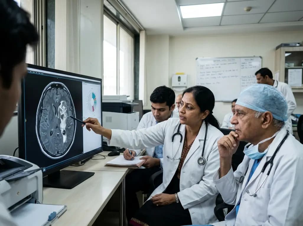
Glioblastoma isocitrate dehydrogenase (IDH)-wildtype represents the most common and aggressive primary central nervous system malignancy in adult neuro-oncology. In recent years, updated classification guidelines have shifted the diagnostic paradigm toward integrated molecular and histological parameters. However, clinical phenotypes still demonstrate substantial heterogeneity across diverse patient populations. Intratumoral hemorrhage represents a distinctive phenotypic manifestation that frequently complicates preoperative radiological assessment and surgical management. Patients presenting with hemorrhagic glioblastoma often experience acute neurological decompensation, which demands rapid diagnostic evaluation and neurosurgical intervention. Despite its striking appearance on neuroimaging, the precise molecular drivers and clinical consequences of intratumoral hemorrhage have remained poorly understood. Researchers have sought to identify whether bleeding within the tumor parenchyma signifies a genetically distinct subclass of IDH-wildtype glioblastoma. Investigating these molecular features is vital because targeted therapies increasingly depend on specific genomic variations. A recent comprehensive genomic investigation explored how hemorrhagic phenotypes diverge from non-hemorrhagic tumors. Understanding these genomic patterns helps clinicians refine prognostic assessments and tailor follow-up strategies. Consequently, illuminating the biological mechanisms underlying hemorrhage provides clinicians with essential insights into intracranial tumor biology and patient care.
Preoperative magnetic resonance imaging (MRI) plays an indispensable role in delineating the structural and hemorrhagic characteristics of high-grade gliomas. To evaluate hemorrhagic patterns objectively, investigators utilized standardized imaging criteria known as Visually Accessible Rembrandt Images (VASARI). In a well-characterized cohort of 176 patients with IDH-wildtype glioblastoma, 105 individuals demonstrated definitive radiological evidence of intratumoral hemorrhage. Clinically, patients with hemorrhagic glioblastoma presented with significantly higher rates of acute focal motor deficits compared to those without hemorrhage. Specifically, motor impairment was documented in 38.0% of hemorrhagic cases compared to only 21.1% in non-hemorrhagic presentations. Furthermore, hemorrhagic glioblastoma lesions exhibited substantially greater total volumes on preoperative imaging, measuring an average of 147.6 cubic centimeters compared to 122.2 cubic centimeters in non-hemorrhagic cases. Demographic comparisons also revealed a significantly higher proportion of Hispanic patients within the hemorrhagic group. These clinical and volumetric disparities highlight the acute mass effect and vascular disruption caused by spontaneous bleeding within the neoplastic tissue. Therefore, neurosurgeons and neuro-oncologists must anticipate substantial edema and sudden functional deterioration when managing patients who present with prominent intratumoral hematomas.
To delineate the molecular architecture of hemorrhagic glioblastoma, investigators performed next-generation sequencing across a targeted panel of 205 cancer-associated genes. This comprehensive genetic analysis revealed several intriguing differences between hemorrhagic and non-hemorrhagic tumor cohorts. Specifically, tumors with intratumoral hemorrhage displayed lower unadjusted alteration frequencies in several key tyrosine kinase and signaling genes. For instance, PDGFRA alterations occurred in only 13.3% of hemorrhagic tumors compared to 26.8% in non-hemorrhagic tumors. Similarly, alterations in KIT (8.5% vs. 19.7%), KDR (6.6% vs. 18.3%), and PIK3R1 (2.8% vs. 11.3%) appeared less frequently in hemorrhagic glioblastoma samples. In contrast, mutations or alterations involving the epigenetic modifier SETD2 occurred with greater frequency in hemorrhagic tumors (8.5% vs. 1.4%). However, after applying rigorous statistical corrections for multiple testing using the Benjamini-Hochberg method, these differences did not maintain formal statistical significance. Nevertheless, these observed biological trends suggest subtle divergence in receptor tyrosine kinase signaling and chromatin regulation. Consequently, these findings indicate that distinct angiogenic and genomic pathways may govern microvascular fragility within aggressive IDH-wildtype tumors.
Determining whether intratumoral hemorrhage confers an inferior clinical prognosis has long been a central question in neuro-oncology. Historically, acute neurological presentations and larger tumor burdens raised concerns about expedited disease progression and decreased survival. However, detailed univariable and multivariable survival analyses from this multi-gene sequencing cohort showed no significant survival penalty associated with hemorrhagic status. In particular, the median progression-free survival was 8.2 months for patients with hemorrhagic glioblastoma versus 8.3 months for those with non-hemorrhagic disease. Similarly, median overall survival reached 18.4 months in the hemorrhagic group compared to 19.7 months in the non-hemorrhagic cohort. These differences were statistically non-significant, demonstrating that the presence of intratumoral hemorrhage does not independently dictate post-surgical survival outcomes. Standard aggressive multimodal management—comprising maximal safe surgical resection, concurrent chemoradiotherapy with temozolomide, and maintenance therapy—remains effective across both radiographic subgroups. As a result, neuro-oncology multidisciplinary teams should avoid assuming a worse oncological trajectory solely based on the radiographic appearance of acute or subacute intratumoral hemorrhage.
The identification of hemorrhagic features introduces distinct technical and perioperative challenges during clinical neurosurgical care. Because hemorrhagic glioblastoma lesions tend to be larger and present with motor weakness, surgical teams must prioritize rapid stabilization, intracranial pressure control, and precise surgical planning. Intraoperatively, extensive vascular dysmorphism, friable neoplastic vasculature, and organized hematomas require meticulous microdissection and rigorous hemostasis. Furthermore, operating neurosurgeons must carefully differentiate between primary intratumoral bleeding and non-neoplastic vascular malformations or hypertensive intracerebral hemorrhage during emergency craniotomies. Postoperatively, multimodal therapy should proceed without unnecessary delays once surgical wounds have stabilized. Although targeted agents directed against receptor tyrosine kinases like PDGFRA or KDR are currently explored in clinical trials, the lower frequency of these mutations in hemorrhagic glioblastoma emphasizes the need for individualized genetic profiling. Clinicians must rely on comprehensive molecular panels rather than phenotypic assumptions when evaluating eligibility for targeted therapeutic protocols. Therefore, coordinated care involving neurosurgeons, neuroradiologists, neuropathologists, and medical oncologists remains paramount for achieving optimal patient outcomes.
While current retrospective data clarify overall survival equivalence, further translational studies are needed to uncover the exact pathophysiological mechanisms driving vascular instability. Future investigations should incorporate multi-omic profiling, including single-cell transcriptomics, spatial transcriptomics, and epigenetic analyses, to characterize the tumor microenvironment surrounding intratumoral hematomas. In addition, prospective multi-center registries can help elucidate why certain demographic subsets, such as Hispanic populations, demonstrate a higher prevalence of hemorrhagic presentation. Researchers should also explore advanced radiomic and artificial intelligence models to predict microvascular fragility before overt bleeding occurs. Such predictive imaging biomarkers could inform perioperative anticoagulation protocols and venous thromboembolism management, which frequently complicate glioblastoma care. Moreover, expanding molecular panels beyond 205 genes might reveal novel driver mutations responsible for vascular friability and altered endothelial integrity. Ultimately, integrating molecular genomics with high-resolution neuroimaging will continue to refine risk stratification and therapeutic development for all glioblastoma phenotypes.
Hemorrhagic glioblastoma is defined by the presence of acute, subacute, or chronic intratumoral bleeding visible on MRI sequences. Radiologists routinely employ standardized frameworks, such as the Visually Accessible Rembrandt Images criteria, to identify parenchymal hemorrhage, internal blood-fluid levels, and associated mass effect within high-grade gliomas.
Current genomic and clinical evidence demonstrates that intratumoral hemorrhage does not significantly worsen progression-free or overall survival in IDH-wildtype glioblastoma. Patients receiving standard maximal safe resection and multimodal chemoradiotherapy achieve survival outcomes comparable to individuals diagnosed with non-hemorrhagic glioblastoma phenotypes.
Investigators analyzed 205 cancer-related genes using comprehensive next-generation sequencing. They observed unadjusted differences in PDGFRA, KIT, KDR, PIK3R1, and SETD2 alteration frequencies between hemorrhagic and non-hemorrhagic tumors, although these variations did not remain statistically significant following multiple testing corrections.
Disclaimer: This content is for informational and educational purposes only and is intended solely for healthcare professionals. It should not be used as a substitute for professional medical advice, diagnosis, or treatment. Refer to the latest local and national guidelines for clinical practice.
References

Read summarized clinical updates, watch expert medical content, and earn CME certifications right from your smartphone.
Open your smartphone camera to scan and install.





A retrospective analysis of IDH-wildtype glioblastoma evaluated the molecular and clinical profile of hemorrhagic glioblastoma. While hemorrhagic tumors were larger and presented with motor deficits, next-generation sequencing and survival analyses revealed comparable overall survival and progression-free survival.
last month

UK Research and Innovation is deploying £80 million across the NHS to trial nine cutting-edge technologies. The initiative features seven-minute brain MRIs, blood biomarker assays, and predictive AI tools, aiming to evaluate 92% of suspected cases within 18 weeks and overcome lengthy diagnostic backlogs.
Today

A mixed-methods study identifies key drivers of PMTCT service utilization among adolescent girls and young women aged 15-24. Highlighting age disparities, PrEP uptake, and systemic barriers, the evidence guides clinicians toward adolescent-responsive, integrated maternal HIV prevention protocols.
Today

PepsiCo and Monster Beverage have challenged the FSSAI ban on the energy drink label in Indian courts. While companies highlight supply disruptions, regulatory actions spotlight severe cardiovascular and metabolic risks associated with excessive caffeine, sugar, and taurine consumption, especially in adolescents.
Today

A narrative review investigates whether the articularis genus muscle functions as an independent anatomical entity or blends with the vastus intermedius. We evaluate its morphology, role in retracting the suprapatellar bursa, and direct clinical significance in anterior knee pain and arthroplasty.
Today

A Bayesian multilevel meta-analysis reveals that aerobic training combined with moderate carbohydrate restriction modestly lowers HbA1c in type 2 diabetes. However, sparse data and very low certainty leave incremental benefits over exercise or diet alone unproven, highlighting the need for individualized care.
Yesterday
