.webp&w=384&q=75)

Loading, please wait...
Loading, please wait...
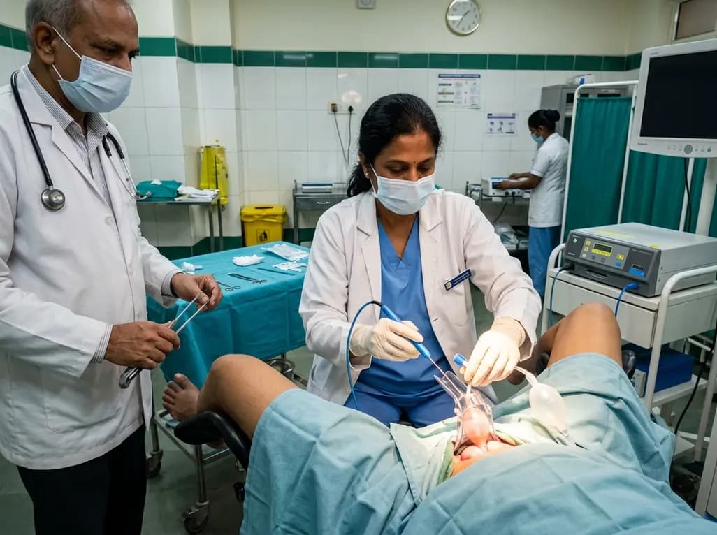
Cervical intraepithelial neoplasia represents a significant clinical challenge in preventive women's healthcare worldwide. Loop electrosurgical excision procedure remains the standard outpatient surgical modality for diagnosing and treating high-grade cervical dysplasia. However, technical difficulties such as anatomical mobility, inadequate exposure, and tissue collapse often complicate the excision pass. Consequently, clinicians frequently encounter fragmented specimens that compromise histopathological margin evaluation. To address these operative barriers, researchers have introduced a modified LEEP technique utilizing gentle Foley catheter balloon traction. This innovative surgical refinement aims to stabilize the ectocervix, maintain anatomical orientation, and facilitate a continuous single-pass excision without disrupting baseline surgical efficiency or patient safety.
Precise surgical geometry during cervical conization directly dictates clinical success. When a surgeon performs an electrosurgical loop excision, maintaining steady counter-traction is essential. Traditional techniques often rely on single-tooth tenacula or simple tissue forceps. Unfortunately, these instruments can cause focal cervical tearing, bleeding, or premature thermal disruption when the loop nears the anchoring site. Moreover, patient movement or anatomical variations such as pelvic organ prolapse, vaginal wall laxity, or a flush cervix can obscure the transformation zone. Inadequate stabilization frequently forces the operating surgeon to perform multiple fragmented passes instead of a single, uniform tissue sweep. In turn, specimen fragmentation introduces severe challenges during microscopic analysis. When pathologists receive fragmented biopsies, determining true margin involvement becomes exceptionally complex. Fragmented edges produce artificial surgical margins and increase thermal artifact distortion. Therefore, securing stable cervical traction without creating traumatic lacerations is crucial for achieving high-quality specimens, complete lesion removal, and accurate diagnostic staging.
The modified LEEP technique integrates an accessible, cost-effective tool to establish dynamic cervical traction. During this refined procedure, the clinician introduces a small-caliber Foley catheter through the external cervical os into the lower uterine canal before applying electrosurgical energy. Once positioned, the balloon is gently inflated with sterile saline to create an internal anchor against the internal os. By applying mild, coaxial outward traction on the catheter shaft, the surgeon effortlessly draws the cervix into an optimal focal plane. Furthermore, this coaxial tension symmetrically flattens the ectocervical transformation zone. As a result, the surgeon gains an unobstructed, panoramic view of the entire lesion boundary. The electrosurgical wire loop can then glide seamlessly beneath the transformation zone in a smooth, continuous arch around the catheter. Because the traction force originates centrally from the endocervical canal rather than from peripheral grasping instruments, the perimeter remains entirely clear for clean margin clearance.
Recent clinical evidence highlights significant histopathological advantages associated with this catheter-assisted approach. In a retrospective feasibility study analyzing 108 patients undergoing excisional therapy, the modified technique demonstrated zero instances of specimen fragmentation. Conversely, standard loop excision cohorts exhibited multiple fragmented specimens requiring complex histological reconstruction. Pathologists consistently reported that intact single-specimen cones enabled precise radial inking and definitive evaluation of both ectocervical and endocervical margins. Furthermore, overall margin positivity rates remained comparable between the modified and standard surgical cohorts. This finding confirms that the catheter balloon does not push the lesion outward or cause artificial margin compromise. Histopathologists emphasize that an intact cone specimen minimizes inconclusive margin reports, which directly reduces patient anxiety and prevents unnecessary follow-up interventions. Achieving clean, evaluable specimens represents a vital quality benchmark in modern gynecologic oncology.
Introducing any surgical modification demands rigorous evaluation of operating time, complication rates, and perioperative safety. Current data confirm that the catheter traction method is both reproducible and straightforward to master. Operating room setup times remain practically identical to conventional protocols, requiring only standard clinic consumables. Importantly, the modified approach does not increase intraoperative bleeding or postoperative hemorrhage risks. Because the inflated balloon provides mild internal tamponade along the lower endocervical canal, localized vascular oozing often diminishes during initial tissue handling. In addition, postoperative infection rates, healing timelines, and short-term recovery profiles mirror those of standard loop procedures. Surgeons also report superior ergonomics, as the steady traction reduces the need for awkward speculum adjustments or awkward assistant retraction. Consequently, this refined method offers a safe, accessible adaptation that easily integrates into routine outpatient surgical workflows without demanding expensive specialized hardware.
The integration of balloon-assisted traction offers widespread clinical utility across diverse healthcare settings. In high-volume colposcopy clinics and low-resource environments, optimizing the diagnostic yield of every single procedure is essential. Cervical dysplasia screening programs rely heavily on accurate histology to guide surveillance intervals and avoid repeat conizations. By preventing specimen fragmentation, this novel modification enhances diagnostic confidence and streamlines subsequent patient management. Furthermore, the standardized exposure created by symmetrical traction makes the procedure exceptionally valuable for resident training and surgical education. Novice clinicians can better visualize the depth and lateral margins of the wire loop pass. While these preliminary findings provide compelling proof-of-concept data, prospective randomized multicenter trials should follow. Future studies will fully quantify long-term cervical competency, stenosis rates, and oncologic recurrence outcomes across larger, diverse patient populations.
The inflated catheter balloon anchors gently within the endocervical canal, allowing the surgeon to apply mild outward traction. This maneuver draws a recessed or mobile cervix forward into direct view, flattening the transformation zone symmetrically. Consequently, the clinician achieves an unobstructed field, eliminating vaginal wall interference and facilitating a clean, single-pass loop excision without peripheral instrument obstruction.
Current clinical findings show no increased incidence of cervical stenosis or canal scarring compared to standard excision methods. The catheter balloon is inflated with minimal volume only to establish gentle counter-traction, preventing endocervical mucosal trauma. Therefore, the tissue healing process, re-epithelialization timeline, and long-term cervical canal patency remain entirely comparable to conventional loop electrosurgical procedures.
Fragmented specimens severely hinder microscopic analysis because pathologists cannot accurately distinguish true surgical margins from artificial cuts. This ambiguity often leads to indeterminate margin reports and diagnostic uncertainty regarding residual disease. Preserving an intact, single-piece cone ensures clear margin visualization, reduces thermal artifact distortion, and helps clinicians establish definitive post-treatment surveillance or management plans.
Disclaimer: This content is for informational and educational purposes only and does not constitute formal medical advice, diagnosis, or treatment recommendations. Clinical decisions must always rely on individual patient assessments, institutional protocols, and multidisciplinary surgical evaluations. Refer to the latest local and national guidelines for clinical practice.
References

Read summarized clinical updates, watch expert medical content, and earn CME certifications right from your smartphone.
Open your smartphone camera to scan and install.





A novel modified LEEP technique employing Foley catheter traction enhances cervical stabilization and specimen integrity during dysplasia excision, eliminating specimen fragmentation while maintaining comparable margin positivity and safety profiles.
4 weeks back

Red blood cell transfusion is critical in sickle cell disease management but brings significant immunological complexity, including alloimmunisation and iron overload. Extended RBC phenotyping and tailored protocols optimize clinical safety.
Today
Discover the crucial distinctions between mesonephric and mesonephric-like proliferations of the female genital tract. Learn the key histomorphologic, immunohistochemical, and molecular differences essential for accurate diagnosis and clinical management.
Today

A large UK Biobank study reveals that plasma proteomic signatures capture preclinical organ damage and improve multiorgan risk prediction across early CKM syndrome stages 0 to 2 beyond traditional PREVENT clinical models.
Today

A retrospective CBCT study reveals that superior TMJ space asymmetry correlates moderately with occipital condyle-atlas-axis asymmetry in unilateral TMJ osteoarthritis, highlighting an imaging correlate between jaw biomechanics and upper cervical alignment rather than a standalone diagnostic marker.
Today

A Bayesian multilevel meta-analysis reveals that aerobic training combined with moderate carbohydrate restriction modestly lowers HbA1c in type 2 diabetes. However, sparse data and very low certainty leave incremental benefits over exercise or diet alone unproven, highlighting the need for individualized care.
Today
