.webp&w=384&q=75)

Loading, please wait...
Loading, please wait...
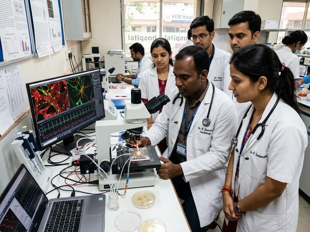
The cardiac autonomic nervous system plays an indispensable role in maintaining cardiovascular homeostasis and regulating chronotropic and inotropic states. However, dysregulation of autonomic signaling represents a major driver of malignant arrhythmias, heart failure, and sudden cardiac death. Historically, investigating human neuro-cardiac interactions has presented major experimental challenges because animal models frequently fail to recapitulate human cardiac electrophysiology and autonomic innervation patterns. Consequently, conventional monoculture systems cannot reproduce the complex spatial and biochemical crosstalk between sympathetic nerves and myocardial tissue. To bridge this translational gap, researchers have developed innovative microphysiological platforms that faithfully reconstitute these cellular communication networks. By combining compartmentalized microfluidic architectures with human induced pluripotent stem cell (hiPSC) technology, investigators can now observe human autonomic regulation in real time. Therefore, this technological paradigm provides clinicians and cardiovascular researchers with unprecedented mechanistic precision to explore how sympathetic hyperactivity triggers arrhythmogenic substrate remodeling.
To accurately recreate the anatomical interface between nerve terminals and myocardial cells, bioengineers implemented a compartmentalized microfluidic device (MFD). Specifically, the platform features two distinct open chambers connected by a series of high-resistance microchannels. This geometric separation permits the selective culturing of sympathetic neurons in one chamber and cardiomyocytes in the adjacent chamber. Furthermore, the microscopic channel dimensions permit directed axonal extension across the physical divide while restricting neuronal cell somas to their compartment. As a result, fluidic isolation allows researchers to apply pharmacological agents or electrical stimulation selectively to neuronal or cardiac compartments without unintended cross-contamination. In addition, the optical clarity of the system facilitates continuous high-resolution live imaging and patch-clamp electrophysiology. This engineered microenvironment successfully mirrors physiological neuro-cardiac architecture, thereby establishing a reproducible in vitro surrogate for advanced translational research.
Establishing an authentic autonomic response requires robust differentiation and comprehensive functional validation of neuronal lineages. In this microphysiological platform, investigators differentiated hiPSCs into mature sympathetic neurons (hiPSC-SNs) confirmed by quantitative molecular and structural assays. Notably, reverse transcription quantitative PCR (RT-qPCR) and immunofluorescence analyses revealed substantial upregulation of signature sympathetic markers, including peripherin, tyrosine hydroxylase, and βIII-tubulin. Moreover, the differentiated neurons exhibited high transcriptional expression of dopamine β-hydroxylase alongside functional nicotinic acetylcholine receptors. Whole-cell patch-clamp electrophysiology further substantiated their functional maturation by capturing spontaneous action potential firing. In addition, localized application of 1 micromolar nicotine triggered rapid membrane depolarization and action potential bursts. Consequently, these multi-parametric findings confirm that the differentiated neurons possess the necessary biochemical machinery and bioelectric properties characteristic of native sympathetic postganglionic fibers.
Physical proximity and directional axonal guidance remain fundamental requirements for establishing functional neuro-cardiac coupling in vitro. Over a 10-day co-culture period, hiPSC-SNs extended dense axonal projections through the intervening microfluidic channels into the cardiomyocyte compartment. Consequently, these regenerating axonal terminals formed intimate structural junctions with adjacent hiPSC-derived cardiomyocytes (hiPSC-CMs). To evaluate whether structural connectivity translated into functional autonomic control, investigators performed synchronized patch-clamp recordings and high-speed video-motion tracking. Interestingly, co-cultured cardiomyocytes demonstrated a baseline increase in spontaneous action potential frequency and mechanical beating velocity compared to uninnervated controls. Thus, baseline tonic signaling from sympathetic axons actively modulates myocardial excitability within the device. These observations confirm the successful establishment of a functional neuro-effector junction that replicates human autonomic physiology.
A major advantage of this microphysiological system lies in its capacity for precise pharmacological interrogation of autonomic signaling pathways. When researchers applied nicotine selectively to the neuronal compartment, the sympathetic axons released neurotransmitters that significantly amplified cardiomyocyte action potential rates and contraction frequencies. Conversely, introducing the non-selective β-adrenergic receptor antagonist propranolol (5 micromolar) abolished this positive chronotropic effect and markedly decreased cardiomyocyte beating rates. In addition, this pharmacological responsiveness proves that signal transduction across the microfluidic channel relies genuinely on adrenergic receptor activation. Therefore, this model provides a scalable testbed for evaluating novel anti-arrhythmic agents, autonomic modulators, and cardiotoxic compounds. Furthermore, because the platform uses human cells, it avoids interspecies discrepancies in adrenergic receptor density and ion channel kinetics, delivering clinically translatable pharmacological data.
Autonomic nervous system dysfunction represents a central pathophysiological mechanism in long QT syndrome, catecholaminergic polymorphic ventricular tachycardia, and ischemic arrhythmias. Importantly, patient-specific hiPSCs carrying pathogenic genetic variants can be integrated directly into this microphysiological architecture. By utilizing patient-derived cells, electrophysiologists can investigate how specific channelopathies or autonomic abnormalities precipitate ventricular tachyarrhythmias under adrenergic stress. Additionally, this platform enables clinicians to test personalized pharmacological regimens in vitro before initiating complex anti-arrhythmic therapies in vulnerable patients. Furthermore, the technology opens new therapeutic vistas for evaluating targeted autonomic neuromodulation therapies, such as renal denervation and stellate ganglion blockade surrogates. Ultimately, this biomimetic approach bridges the gap between basic electrophysiology and bedside clinical cardiology, advancing precision medicine for complex cardiac disorders.
The compartmentalized microfluidic platform utilizes high-resistance microchannels between the wide-open culture chambers. These microchannels establish laminar flow resistance and hydrostatic pressure barriers that prevent bulk fluid exchange between chambers. Consequently, researchers can selectively apply drugs, agonists, or antagonists to sympathetic neuronal somas without non-specific diffusion into the cardiomyocyte compartment, ensuring pure neuro-effector evaluation.
Animal models frequently present distinct electrophysiological profiles, such as divergent resting membrane potentials, action potential durations, and adrenergic receptor subtypes compared to humans. In contrast, human induced pluripotent stem cell models express authentic human ion channel repertoires and autonomic receptor distributions, thereby avoiding interspecies translation failures and providing physiologically relevant data for human drug screening.
Nicotine acts selectively on nicotinic acetylcholine receptors located on sympathetic neuronal cell bodies in the neuronal chamber. Upon stimulation, the neurons depolarize and propagate action potentials along their axons into the cardiac chamber, releasing endogenous catecholamines. This localized release stimulates myocardial β-adrenergic receptors, causing measurable increases in cardiomyocyte beating frequency that are blocked by propranolol.
Disclaimer: This content is for informational and educational purposes only and should not be considered medical advice or a substitute for professional clinical judgment. Diagnostic and treatment decisions must always be tailored to individual patients by qualified healthcare professionals. While based on verified clinical sources, medical knowledge is constantly evolving. Refer to the latest local and national guidelines for clinical practice.
References

Read summarized clinical updates, watch expert medical content, and earn CME certifications right from your smartphone.
Open your smartphone camera to scan and install.





Researchers have engineered a human iPSC-based microphysiological system that successfully couples sympathetic neurons with cardiomyocytes, offering crucial insights into autonomic heart regulation and opening novel avenues for cardiac disease modeling and targeted drug discovery.
last month

A systematic review of 41 preclinical studies reveals that baicalin dampens systemic inflammation, inhibits NF-κB and NLRP3 pathways, and improves organ preservation in sepsis models. However, substantial study heterogeneity and a complete lack of human clinical trials highlight the need for further translational research.
Today
Discover the crucial distinctions between mesonephric and mesonephric-like proliferations of the female genital tract. Learn the key histomorphologic, immunohistochemical, and molecular differences essential for accurate diagnosis and clinical management.
Today

A large UK Biobank study reveals that plasma proteomic signatures capture preclinical organ damage and improve multiorgan risk prediction across early CKM syndrome stages 0 to 2 beyond traditional PREVENT clinical models.
Today
A narrative review investigates whether the articularis genus muscle functions as an independent anatomical entity or blends with the vastus intermedius. We evaluate its morphology, role in retracting the suprapatellar bursa, and direct clinical significance in anterior knee pain and arthroplasty.
Today

A Bayesian multilevel meta-analysis reveals that aerobic training combined with moderate carbohydrate restriction modestly lowers HbA1c in type 2 diabetes. However, sparse data and very low certainty leave incremental benefits over exercise or diet alone unproven, highlighting the need for individualized care.
Today
