.webp&w=384&q=75)

Loading, please wait...
Loading, please wait...
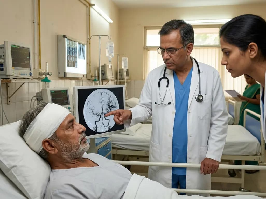
Chronic subdural hematoma represents one of the most common neurosurgical conditions in elderly populations. Surgical evacuation, using burr-hole craniotomy or open formal craniotomy, remains the primary treatment for symptomatic hematomas. However, hematoma recurrence remains a significant clinical challenge. To reduce recurrence rates, middle meningeal artery embolization has emerged as an effective adjunctive endovascular therapy. This procedure targets the fragile hypervascular neomembranes supplying the hematoma cavity. During diagnostic angiography prior to embolization, clinicians occasionally encounter abnormal vascular shunts. Specifically, MMA arteriovenous shunting has been reported after cranial trauma and neurosurgical procedures, yet its exact frequency after chronic subdural hematoma surgery remained poorly defined.
Interventional specialists must recognize these postoperative vascular changes to distinguish transient low-flow shunts from high-risk dural arteriovenous fistulas. Understanding how surgical techniques influence shunt development provides essential guidance for clinical management. This article reviews recent evidence on the frequency, angiographic characteristics, and endovascular management of middle meningeal artery shunts following surgical hematoma evacuation.
To investigate this postoperative vascular phenomenon, researchers conducted a retrospective cohort study evaluating consecutive patients undergoing middle meningeal artery embolization after surgical evacuation of chronic subdural hematoma. The study included thirty consecutive patients treated between January 2020 and March 2023. Each patient underwent selective and superselective middle meningeal artery angiography during the embolization procedure. Experienced neurointerventional specialists systematically reviewed the resulting digital subtraction angiograms.
The investigators analyzed several key angiographic variables, including shunt presence, structural morphology, flow dynamics, early venous drainage, and microcatheter positioning. Surgical procedures were categorized as either burr-hole craniotomy or formal open craniotomy. Additionally, post-embolization control angiography evaluated immediate shunt obliteration following embolic delivery. This methodical approach enabled researchers to characterize shunt dynamics and evaluate the direct impact of surgical exposure on dural vascular integrity. Superselective microcatheter injections provided high-resolution visualization of subtle distal vascular communications that standard diagnostic runs could easily miss.
The study demonstrated a strong correlation between the surgical technique used for hematoma evacuation and the development of post-surgical shunting. Angiographic evidence of MMA arteriovenous shunting was identified in eleven out of thirty patients, establishing an overall incidence of thirty-six point seven percent. This high prevalence indicates that post-surgical vascular communications are far more frequent than previously recognized in routine practice.
When stratified by surgical approach, a substantial disparity was observed between open craniotomy and burr-hole craniotomy. Patients who underwent formal craniotomy exhibited a ninety point nine percent rate of arteriovenous shunting. In contrast, patients treated with burr-hole craniotomy demonstrated a shunting rate of only five point three percent. Statistical testing confirmed that this difference was highly significant. Formal open craniotomy requires extensive dural elevation and vessel disruption, which likely promotes iatrogenic fistula formation. Conversely, burr-hole craniotomy involves minimal mechanical dural disruption, significantly reducing the likelihood of creating abnormal arteriovenous connections.
Comprehensive angiographic assessment provided valuable details regarding shunt architecture and flow dynamics. Most shunts were detectable during initial selective angiography before microcatheter advancement, proving they were not caused by microcatheter manipulation. Superselective microcatheter angiography further refined visualization of delicate shunt morphology and venous pathways.
Importantly, all observed shunts exhibited low-flow angiographic features without evidence of cortical venous reflux. Cortical venous reflux is a high-risk feature associated with intracranial hypertension, venous infarction, and hemorrhage. Because these post-surgical shunts drained exclusively into dural veins or sinus structures, they carried low immediate clinical risk. Recognizing these low-flow characteristics reassures clinicians while preventing misinterpretation. Postoperative shunts can occasionally mimic active contrast extravasation or persistent neomembrane hypervascularity. Accurate identification allows interventionalists to distinguish low-risk dural shunts from active bleeding or malignant fistulas, ensuring safe and appropriate embolic delivery during the endovascular procedure.
Middle meningeal artery embolization proved to be a highly effective treatment for both the subdural hematoma neomembranes and concurrent vascular shunts. The primary objective of embolization is devascularizing hypervascular membranes to prevent recurrent subdural hemorrhage. However, embolic material delivery also directly addresses concurrent arteriovenous communications by occluding feeding arterial branches.
In all eleven affected patients, post-embolization control angiography demonstrated complete disappearance of the arteriovenous shunts. Standard embolic agents successfully closed the fistulous connections without procedural complications. This uniform success confirms that routine middle meningeal artery embolization effectively resolves post-surgical shunting while simultaneously controlling neomembrane hypervascularity. Because all low-flow shunts closed completely upon completion of the procedure, additional targeted fistula interventions or surgical revisions were unnecessary. Immediate post-procedural angiographic confirmation provided definitive proof of complete devascularization, reinforcing the clinical versatility and safety of endovascular embolization in postoperative neurovascular management.
Systematic characterization of postoperative middle meningeal artery shunting provides practical insights for neurosurgeons and interventional radiologists. MMA arteriovenous shunting is a frequent finding after surgical evacuation of chronic subdural hematoma, particularly following formal open craniotomy. Anticipating this phenomenon during postoperative neuroangiography prevents misdiagnosis and avoids unnecessary surgical interventions.
Although the precise long-term clinical impact of untreated low-flow shunts requires further investigation, their consistent resolution during standard embolization offers substantial therapeutic reassurance. Middle meningeal artery embolization successfully achieves dual clinical goals by preventing hematoma recurrence and obliterating iatrogenic vascular shunts. As endovascular embolization becomes increasingly integrated into chronic subdural hematoma care pathways, understanding these benign vascular changes will enhance procedural safety. Neurointerventional teams can confidently proceed with routine embolic penetration, knowing that complete angiographic cure of both the neomembrane and the postoperative shunt is readily achievable in a single session.
MMA arteriovenous shunting occurs primarily due to mechanical surgical trauma to dural blood vessels. During surgical evacuation, particularly open formal craniotomy, elevation of the dural flap and vessel manipulation create small fistulous communications between the middle meningeal artery and adjacent middle meningeal veins.
Surgical technique significantly impacts shunting frequency. Patients undergoing open formal craniotomy experience a much higher incidence of shunting (90.9%) compared to those undergoing minimally invasive burr-hole craniotomy (5.3%). Extensive dural incision and vessel manipulation in formal craniotomy increase the likelihood of iatrogenic arteriovenous fistula formation.
Yes, endovascular middle meningeal artery embolization is highly effective. Post-embolization control angiography demonstrated complete closure and disappearance of MMA arteriovenous shunts in 100% of affected patients. The embolic material successfully obliterates both the hypervascular subdural neomembranes and the low-flow fistulous communications simultaneously during the procedure.
Disclaimer: This content is for informational and educational purposes only... Refer to the latest local and national guidelines for clinical practice.
References

Read summarized clinical updates, watch expert medical content, and earn CME certifications right from your smartphone.
Open your smartphone camera to scan and install.





A retrospective cohort study evaluated MMA arteriovenous shunting in patients after chronic subdural hematoma evacuation. Shunting occurred in 36.7% of patients, with a significantly higher rate after formal craniotomy (90.9%) than burr-hole craniotomy (5.3%). Embolization achieved complete closure in all cases.
2 months ago

As monsoon seasons elevate arboviral disease threats, Tamil Nadu has initiated an ICMR-monitored evaluation of the tetravalent dengue vaccine in Tenkasi district. Clinicians emphasize clinical monitoring, public prevention, and high-quality early pediatric care to reduce disease morbidity across endemic regions.
Today

Leucine Rich Bio has introduced South Asia's first comprehensive vaginal microbiome test, BugSpeaks. By profiling microbial dysbiosis, pathogens, and antimicrobial resistance, this next-generation diagnostic platform provides clinicians with deeper insights into recurrent infections, fertility, and reproductive health.
Today

Review how tailored exercise programs benefit frail older patients with heart failure. Explore pathophysiological mechanisms, individualized training prescriptions, and strategies to overcome adherence barriers in clinical practice.
Today

This comparative study evaluates posterior labral tears with and without glenohumeral instability, highlighting distinct demographic patterns, MRI underestimation in stable shoulders, and successful arthroscopic repair outcomes.
Today

A clinical study demonstrates that early intervention with tocilizumab, either as monotherapy or bridged with corticosteroids, achieves significant proptosis reduction and resolves active inflammation in treatment-naive thyroid eye disease.
Today
