

Loading, please wait...
Loading, please wait...

Managing oral rehabilitation in young children diagnosed with hypohidrotic ectodermal dysplasia requires innovative restorative approaches that accommodate skeletal growth while restoring vital oral functions. Hypohidrotic ectodermal dysplasia typically manifests as severe hypodontia or anodontia, conical anterior teeth, underdeveloped alveolar ridges, and reduced lower facial height. Consequently, young patients suffer from significant masticatory deficiencies, speech impediments, and severe psychosocial distress during formative developmental stages. Traditional pediatric prostheses often fail due to compromised retention, unstable ridge morphology, and the mechanical weakness of conventional acrylic resins. However, advances in computer-aided design and computer-aided manufacturing have transformed modern reconstructive dentistry. By uniting digital workflows with magnetic retention assemblies and monolithic materials, clinicians can now construct highly durable, precise, and easily modifiable removable prostheses. This digital strategy protects developing tissues and supports continuous facial and psychological development in affected children.
Hypohidrotic ectodermal dysplasia represents a rare congenital disorder characterized by the defective development of ectodermal structures, notably hair, sweat glands, and dentition. Because ectodermal differentiation fails during embryogenesis, dental practitioners frequently encounter severe oligodontia or complete anodontia across both primary and permanent dentitions. Furthermore, the few teeth that do emerge frequently present with dysplastic, conical, or peg-shaped crowns supported by short, fragile roots. Underdeveloped alveolar processes invariably accompany this missing dentition because alveolar bone requires dental follicles for vertical and horizontal bone deposition. Therefore, affected children present with sunken perioral soft tissues, prominent lips, and an aged senile facial profile. In addition, reduced salivation often complicates mucosal tolerance, making conventional prosthodontic appliances uncomfortable. Dental clinicians must recognize these clinical signs early in life. Early recognition facilitates timely functional rehabilitation, which safeguards normal speech acquisition, prevents temporomandibular joint dysfunction, and preserves remaining bony structures throughout childhood development.
Prosthetic management of pediatric patients presents unique biological and mechanical dilemmas that differentiate it from adult rehabilitation. Most importantly, continuous transverse and sagittal growth of the maxilla and mandible precludes the early placement of osseointegrated dental implants. Implants act like ankylosed units that arrest local skeletal growth and cause severe positional discrepancies over time. Consequently, clinicians must rely on removable prostheses as the primary restorative option during childhood and early adolescence. However, traditional removable partial dentures feature numerous limitations in young patients. Conventional cold-cured or heat-cured polymethyl methacrylate bases frequently harbor internal porosity, retain residual monomers, and fracture under cyclic masticatory loads. In addition, traditional artificial denture teeth rely solely on chemical bonding to the base, resulting in frequent tooth debonding and urgent repair visits. Repeated mechanical fractures discourage children and compromise compliance. Therefore, restorative protocols require robust materials and adaptable designs that withstand vigorous chewing forces while accommodating routine adjustments.
Digital impression techniques and subtractive computer-aided manufacturing offer significant improvements over conventional laboratory workflows. Clinicians can capture accurate intraoral surface scans without inducing nausea or anxiety in pediatric patients. Following optical capture, specialized design software constructs monolithic prostheses using dual-shade polymethyl methacrylate pucks. Because the entire framework and teeth mill from a single industrial block of prepolymerized resin, no adhesive interface exists between the denture teeth and the pink denture base. Consequently, this monolithic architecture completely eliminates the risk of artificial tooth detachment during chewing. Moreover, prepolymerized resin pucks possess superior mechanical properties, higher flexural strength, and virtually zero residual monomer release compared to conventional materials. Industrial polymerization also provides excellent surface smoothness, which resists bacterial biofilm colonization and plaque accumulation. Thus, digital monolithic milling ensures exceptional structural durability while streamlining future digital modifications as the child grows.
Achieving predictable prosthesis retention presents a major hurdle when alveolar ridges are severely hypoplastic and few conical teeth exist. Traditional clasp assemblies exert detrimental lateral torque forces on malformed abutment teeth with compromised periodontal support. In contrast, closed-field magnetic attachment systems provide passive, predictable vertical retention without applying harmful lateral leverage to fragile abutment roots. Clinicians first restore the conical natural abutments using milled prepolymerized resin crowns designed with precise internal recesses for magnetic keepers. Corresponding reciprocal recesses are digitally planned into the intaglio surface of the monolithic removable partial denture. As a result, the magnetic elements seat completely passively without generating mechanical stress on the supporting teeth during insertion or mastication. In addition, magnetic systems allow straightforward insertion and removal for young patients with developing motor coordination. This atraumatic retention mechanism protects natural tooth structures while delivering outstanding prosthetic stability.
Longitudinal evaluation demonstrates that digital monolithic removable prostheses retained with magnetic attachments provide outstanding clinical outcomes in young patients. At six-month clinical follow-up examinations, patients demonstrate stable occlusion, healthy periodontal tissues around crowned abutments, and uncompromised mucosal integrity beneath the milled denture base. Patients and parents consistently report substantial improvements in chewing efficiency, clearer articulation, and heightened self-confidence in social environments. Nevertheless, clinicians must schedule rigorous recall visits every three to six months to monitor ongoing craniofacial development. Because jaw growth alters occlusal relationships and mucosal adaptation, the dental team can easily modify the digital designs or rebase the monolithic appliances when necessary. Ultimately, this reversible and conservative protocol preserves existing alveolar bone and natural abutments until the patient completes skeletal maturation, at which point permanent implant-supported reconstructions become clinically viable.
Clinicians avoid placing dental implants in growing children because implants lack a periodontal ligament and behave like ankylosed teeth. As the surrounding maxillary and mandibular bones expand through normal skeletal development, the stationary implant fails to migrate with the growing alveolus. This discrepancy leads to severe vertical infraocclusion, altered maxillomandibular relationships, and permanent aesthetic defects. Therefore, practitioners postpone implant placement until skeletal maturation completes.
Monolithic milling shapes the denture base and artificial teeth as a single cohesive unit from a single prepolymerized polymethyl methacrylate puck. Traditional dentures depend on chemical bonding agents between distinct acrylic teeth and the pink base, creating weak interfaces prone to fracture. Monolithic milling completely eliminates this adhesive boundary, thereby preventing tooth detachment and providing superior resistance against heavy cyclic masticatory forces.
Magnetic attachments deliver constant vertical retention while eliminating detrimental lateral tipping forces on fragile, conical abutment teeth. Traditional metal clasps exert dangerous torque on teeth with compromised root length during mastication and appliance removal. In contrast, magnets release when lateral stresses exceed threshold values, shielding the supporting periodontal ligament from destructive leverage while remaining exceptionally easy for young children to insert.
Disclaimer: This content is for informational and educational purposes only and does not constitute medical advice. Healthcare professionals should make clinical decisions based on their independent judgment and individual patient circumstances. Refer to the latest local and national guidelines for clinical practice.
References
Bing Z et al. Monolithic dual-shade milled removable partial denture with magnetic attachments for a pediatric patient with hypohidrotic ectodermal dysplasia: A clinical report. J Prosthet Dent. 2026 Nov 07. doi: undefined. PMID: 42998942.
Mittal M, Srivastava D, Kumar A, Sharma P. Dental management of hypohidrotic ectodermal dysplasia: A report of two cases. Contemp Clin Dent. 2015;6(3):414-417.
Sinada N, Wang CI. Fixed prosthodontic rehabilitation for an adolescent patient with ectodermal dysplasia using a fully digital workflow: Two-year follow-up. J Prosthet Dent. 2024;131(6):990-995.
Trivedi BD, Bhatia R. Complete and Removable Partial Prosthesis for a Child with Hypohidrotic Ectodermal Dysplasia. Int J Clin Pediatr Dent. 2013;6(1):71-74.

Read summarized clinical updates, watch expert medical content, and earn CME certifications right from your smartphone.
Open your smartphone camera to scan and install.





Explore an innovative digital workflow utilizing monolithic dual-shade milled removable partial dentures and magnetic attachments to treat pediatric hypohidrotic ectodermal dysplasia, improving esthetics and oral function.
Today

Managing cognitive impairment in older cancer patients requires distinguishing delirium, pre-existing dementia, and cancer-related cognitive decline. This comprehensive clinical guide outlines validated screening tools, diagnostic pathways, and decision-making capacity assessments in geriatric oncology.
Today
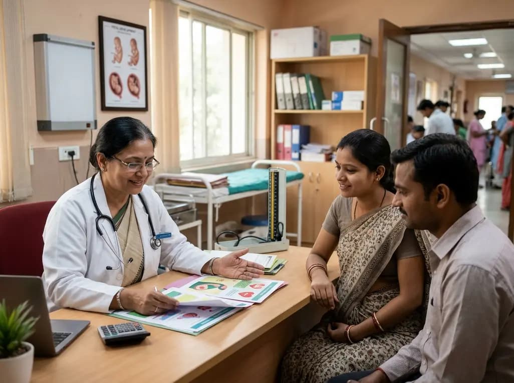
Maternal cardiovascular risk factors such as hypertension, obesity, and diabetes significantly elevate offspring cardiovascular disease risk through placental dysfunction and fetal programming. Enhancing preconception screening and antenatal care offers clinicians a powerful strategy to protect long-term heart health.
Today
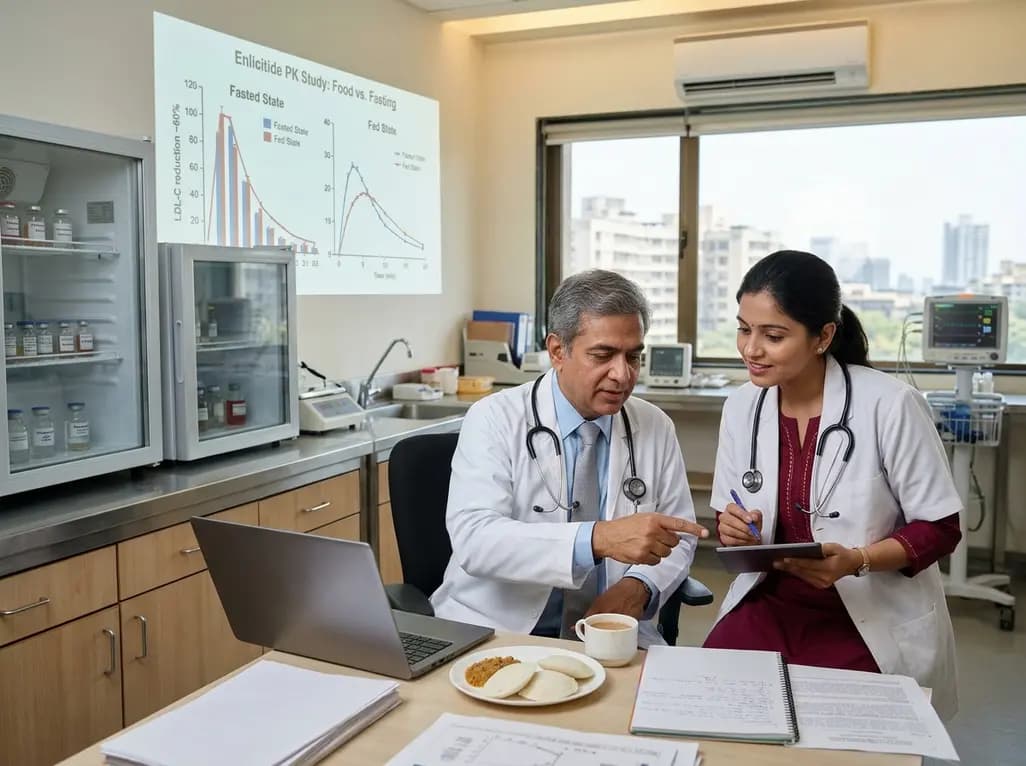
New phase I data evaluate the impact of food and beverages on enlicitide, the first oral PCSK9 inhibitor. While food reduces peak absorption, model simulations demonstrate robust LDL-C lowering of approximately 60%, confirming meaningful pharmacokinetic forgiveness and flexible real-world administration.
Yesterday
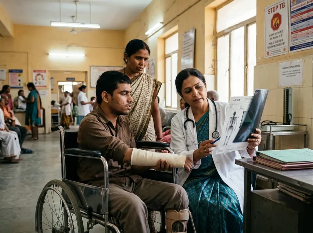
A retrospective cohort study reveals that lumbar spine bone mineral density does not distinguish extremity long bone fracture status in adults with severe motor and intellectual disabilities. Because age drove fracture risk rather than spinal density, clinicians must adopt comprehensive assessment tools.
Today
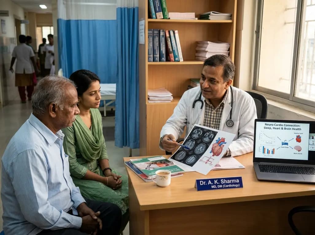
A landmark UK Biobank study by Karolinska Institutet researchers reveals that optimal rest mitigates neurodegeneration in cardiometabolic illness. Patients maintaining high sleep quality demonstrated superior cognitive scores and larger brain volumes, highlighting sleep optimization as an actionable clinical strategy.
Yesterday
