.webp&w=384&q=75)

Loading, please wait...
Loading, please wait...
Mid-aortic syndrome is an uncommon vascular disorder characterized by segmental narrowing of the distal thoracic and abdominal aorta. The condition often involves the visceral and renal arteries, which leads to severe, refractory renovascular hypertension in children and young adults. Because untreated mid-aortic syndrome carries significant morbidity, timely diagnostic identification and effective anatomical revascularization remain paramount for organ preservation and blood pressure control.
Patients with severe aortic narrowing typically present during childhood or adolescence with refractory arterial hypertension. In addition, physical examination often reveals diminished lower-extremity pulses, upper-to-lower limb blood pressure differentials, and continuous abdominal bruits. If clinicians miss these early vascular signs, persistent high pressures can trigger hypertensive encephalopathy, heart failure, or accelerated renal damage. Therefore, vascular specialists utilize contrast-enhanced computed tomography angiography and magnetic resonance angiography to define the coarctation length, luminal caliber, and branch-vessel stenosis.
A thorough diagnostic workup must also differentiate congenital developmental failure from acquired inflammatory diseases such as Takayasu arteritis, neurofibromatosis type 1, or fibromuscular dysplasia. Consequently, clinicians must evaluate inflammatory markers and systemic features before selecting an intervention. Determining whether active vascular inflammation exists helps prevent early graft failure and guides the timing of surgical reconstruction.
Interventional cardiologists and radiologists frequently attempt catheter-based solutions as initial therapies for focal aortic stenoses. However, endovascular interventions face distinct physiological limitations in young patients with complex viscero-renal coarctation. Elastic vessel recoil, severe ostial branching lesions, and vessel rigidity often limit the effectiveness of standard percutaneous transluminal angioplasty. Furthermore, balloon-expandable stents placed across growing aortic segments can complicate future surgical reconstructions.
Consequently, when catheter interventions fail or provide inadequate luminal gain, surgeons must pursue open thoracoabdominal aortic repair. While endovascular stenting may temporize select emergency presentations, open surgical reconstruction remains the definitive benchmark. Open surgery directly establishes adequate downstream perfusion, eliminates the pressure gradient across the stenosis, and preserves long-term viscero-renal parenchymal vitality.
Open surgical repair of thoracoabdominal narrowing demands precise technical execution and customized anatomical planning. Surgeons frequently perform an aorto-aortic bypass or interposition tube graft to bridge the coarcted segment. However, revascularizing the visceral and renal arteries presents substantial technical nuance. In some primary reconstructions, surgeons incorporate the celiac trunk, superior mesenteric artery, and renal arteries as an aortic patch reimplantation into the main prosthetic tube graft.
Nevertheless, severe intrinsic wall rigidity and intimal remodeling within diseased branch ostia can restrict patch inflow. Therefore, when patch reimplantation yields inadequate flow, surgeons must promptly revise the reconstruction by constructing separate prosthetic bypasses to the visceral and renal branches. Creating dedicated individual bypasses often delivers superior hemodynamics and avoids the geometric distortion commonly observed in diseased patch configurations.
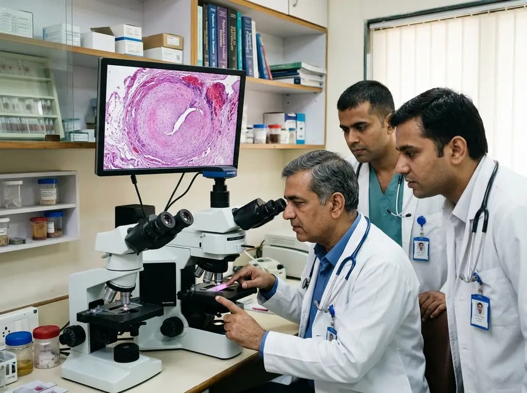
Microscopic tissue analysis of resected aortic segments provides critical insights into the pathophysiology driving luminal compromise. Histopathology in mid-aortic syndrome often reveals prominent adventitial necrosis, smooth muscle disorganization in the media, and severe concentric intimal thickening. These cellular changes confirm that profound luminal compromise results from both structural hypoplasia and exuberant fibrointimal proliferation.
Moreover, extensive intimal fibrosis and medial elastolysis reduce vascular compliance, which directly explains why patch angioplasty and balloon dilatation frequently fail. Recognizing this severe transmural remodeling reinforces the clinical rationale for bypass grafting into healthier, non-stenotic distal targets. Pathological evaluation also helps confirm whether an inflammatory vasculitis drove the underlying destructive mural changes.
Following comprehensive surgical repair, patients usually experience immediate reduction in central arterial pressure and restored distal perfusion. Nevertheless, long-term vascular patency requires vigilant surveillance. Over time, individual bypass grafts—particularly renal revascularization conduits—may undergo late fibrotic occlusion or anastomotic narrowing. Despite unilateral graft occlusion, overall systemic blood pressure and contralateral renal function can remain stable with tailored medical therapy.
Consequently, lifelong multidisciplinary follow-up involving pediatric nephrologists, cardiologists, and vascular surgeons is essential. Serial duplex ultrasound and periodic cross-sectional imaging allow early detection of anastomotic pseudoaneurysms, graft kinking, or progression in native vessels. Dedicated medical management alongside structural surveillance ensures sustained cardiovascular health and preserves end-organ function.
Mid-aortic syndrome can stem from congenital developmental abnormalities, including incomplete fusion of embryonic dorsal aortas, or acquired inflammatory conditions. Genetic syndromes such as neurofibromatosis type 1 and Williams syndrome frequently feature aortic coarctation. Additionally, acquired etiologies like Takayasu arteritis cause severe inflammatory stenosis. Identifying the underlying driver helps clinicians optimize both medical anti-inflammatory therapy and surgical timing.
Endovascular procedures frequently fail because severe intrinsic wall fibrosis, arterial hypoplasia, and extreme elastic recoil resist lasting balloon expansion. Furthermore, significant viscero-renal ostial involvement creates complex branching geometry that stenting cannot reliably correct without compromising vital branch perfusion. In growing pediatric and adolescent patients, rigid metal stents also cannot adapt to ongoing vascular growth.
Histopathological analysis reveals transmural architectural disruption, including adventitial necrosis, medial smooth muscle disarray, and dense intimal hyperplasia. Because this intense fibrosis impairs vessel compliance and compromises patch reimplantation patency, surgeons achieve better long-term durability by constructing separate bypass grafts directly to healthier, disease-free distal vessel segments rather than relying on diseased patch tissue.
Disclaimer: This content is for informational and educational purposes only and is not intended as medical advice. Healthcare professionals should exercise their independent clinical judgment. Refer to the latest local and national guidelines for clinical practice.
References

Read summarized clinical updates, watch expert medical content, and earn CME certifications right from your smartphone.
Open your smartphone camera to scan and install.





Mid-aortic syndrome is a rare vascular condition that causes refractory hypertension in young patients. This report evaluates surgical strategies, bypass revisions, and crucial histopathologic features including adventitial necrosis and intimal hyperplasia that guide clinical decision-making.
4 weeks back

Targeting ferroptosis provides a compelling strategy to dismantle hypoxia- and acidosis-driven immunosuppression in solid tumors, enhancing response to modern immunotherapy.
Today

Findings from Project Viva indicate that sustained postpartum weight retention, rather than excessive gestational weight gain alone, directly worsens women's mid-life cardiovascular health, degrading blood pressure and glycemic parameters decades after delivery.
Today

A large UK Biobank study reveals that plasma proteomic signatures capture preclinical organ damage and improve multiorgan risk prediction across early CKM syndrome stages 0 to 2 beyond traditional PREVENT clinical models.
Today

A retrospective CBCT study reveals that superior TMJ space asymmetry correlates moderately with occipital condyle-atlas-axis asymmetry in unilateral TMJ osteoarthritis, highlighting an imaging correlate between jaw biomechanics and upper cervical alignment rather than a standalone diagnostic marker.
Today

A Bayesian multilevel meta-analysis reveals that aerobic training combined with moderate carbohydrate restriction modestly lowers HbA1c in type 2 diabetes. However, sparse data and very low certainty leave incremental benefits over exercise or diet alone unproven, highlighting the need for individualized care.
Today
