

Loading, please wait...
Loading, please wait...

Surgical intervention within the mesial temporal region demands precise anatomical mastery and refined microsurgical skills. For decades, neurosurgeons have navigated complex vascular networks and eloquent neural tracts to treat drug-resistant temporal lobe epilepsy and structural lesions. Consequently, mesial temporal lobe surgery remains one of the most challenging procedures in modern neurosurgery. To optimize patient outcomes, modern techniques increasingly favor minimally invasive corridors over extensive open resections. A landmark study spanning 26 years provides critical long-term evidence regarding the efficacy and safety of a keyhole craniotomy using an inferior temporal gyrus (ITG) access corridor. By analyzing operative outcomes in 717 consecutive patients, this extensive single-surgeon experience illustrates how targeted trajectories reduce tissue disruption while preserving clinical efficacy. Furthermore, the findings offer practical guidance for both experienced neurosurgeons and trainees navigating deep temporal anatomy.
Historically, neurosurgeons utilized extensive pterional or standard anterior temporal lobectomies to access mesial basal temporal structures. However, these traditional approaches frequently required significant temporal lobe retraction and extensive cortical resection. In contrast, the keyhole craniotomy directed through the inferior temporal gyrus provides an anatomical corridor that minimizes collateral brain disruption. Surgeons place a small, tailored craniotomy centered over the inferior temporal surface. Subsequently, they establish a direct transcortical trajectory through the non-eloquent tissue of the inferior temporal gyrus to reach the temporal horn of the lateral ventricle. This selective trajectory provides immediate visualization of the collateral sulcus, parahippocampal gyrus, and hippocampus. Additionally, the approach avoids the primary language pathways located within the superior and middle temporal gyri. Therefore, surgeons preserve essential neocortical functions while achieving comprehensive mesial cytoreduction. Most importantly, the keyhole approach minimizes brain retraction by exploiting natural anatomical planes, which directly lowers the risk of regional cortical contusion and postoperative cerebral edema.
Understanding patient selection remains crucial when evaluating long-term surgical success. In this 26-year study, investigators evaluated 717 patients undergoing mesial temporal lobe surgery for structural and epileptogenic pathologies. The study cohort had a mean patient age of 37 years, representing a prime working-age demographic where preserving functional independence is paramount. Resection of mesial structures served as the primary surgical objective for all included individuals. However, the study protocol explicitly excluded patients diagnosed with high-grade gliomas showing extensive temporal extension. Because malignant gliomas carry heavy systemic comorbidities and require intensive adjuvant therapy, their exclusion preserved cohort homogeneity. Consequently, the analysis focused cleanly on the intrinsic technical safety of the ITG corridor. The surgical indications primarily comprised hippocampal sclerosis, low-grade neoplasms, vascular malformations, and other focal epileptogenic pathologies. Overall, strict patient selection ensured that surgical outcomes reflected true procedural morbidity rather than baseline oncological decline.
The primary finding of this 26-year review centers on an impressively low complication rate. Among the 717 treated individuals, only 38 patients experienced any perioperative complication, representing an overall rate of 5.3%. Furthermore, researchers divided these events into nonneurological and neurological categories to track clinical trends accurately. Nonneurological complications included superficial surgical site infections in five patients, cerebrospinal fluid leaks or pseudomeningoceles in four patients, and cosmetic defects such as temporalis muscle atrophy in four patients. Additionally, extraaxial hemorrhages developed in eleven patients, representing the single largest nonneurological complication subgroup. Neurological events included transient word-finding difficulties in one patient, transient cranial nerve deficits in two patients, and early postoperative seizures in nine patients. Most remarkably, permanent neurological deficits occurred in only two patients, representing a permanent morbidity rate of just 0.3%. These two permanent deficits comprised one visual field deficit and one lacunar stroke. Importantly, zero perioperative deaths occurred throughout the entire 26-year timeframe.
Achieving a permanent deficit rate of only 0.3% requires meticulous microsurgical strategies designed to prevent neurovascular trauma. For instance, avoiding visual pathway disruption demands strict adherence to anatomical landmarks along the temporal horn roof. Meyer’s loop travels across the anterior and lateral roof of the temporal horn; therefore, entering low through the inferior temporal gyrus effectively skirts below these optic radiation fibers. This deliberate trajectory prevented visual field defects in 99.7% of the cohort. Similarly, protecting critical perforating vessels prevents debilitating ischemic strokes. During resection of the uncus and hippocampal head, surgeons must carefully identify and preserve the anterior choroidal artery and posterior cerebral artery branches. Direct manipulation of the choroidal fissure must proceed with micro-dissectors under high optical magnification. In addition, the surgical team implemented rigorous perioperative antiepileptic management, which minimized acute postoperative seizure exacerbations. Through these standardized technical adjustments, surgical teams successfully neutralized the primary mechanisms driving permanent operative morbidity.
Beyond individual technical steps, this 26-year series illustrates the critical maturation curve of a refined neurosurgical approach. Mesial temporal resections frequently intimidate junior neurosurgeons due to the proximity of the brainstem, internal carotid artery, and cranial nerves. However, this study demonstrates that standardizing a reproducible corridor fosters safe surgical education. Over the multi-decade experience, the senior surgeon systematically incorporated structured operative milestones for assisting fellows and residents. By breaking the procedure into discrete phases—craniotomy placement, ventricular entry, amygdala debulking, and hippocampal resection—trainees acquired procedural mastery safely. In addition, careful post-case debriefs and longitudinal complication tracking directly influenced operative teaching protocols. Consequently, surgical complication rates declined steadily over time despite the active training of junior clinicians. This progressive mastery demonstrates that complex keyhole procedures can safely enter surgical training curricula when senior mentors apply structured anatomical guidelines.
These surgical insights hold immense relevance for neurosurgical practice across India, where drug-resistant epilepsy represents a major healthcare challenge. Many tertiary hospitals in urban and semi-urban settings treat substantial volumes of mesial temporal lobe epilepsy. However, open procedures often encounter reluctance due to patient concerns regarding cosmetic deformities and lengthy recovery times. Adopting a keyhole ITG corridor addresses these hurdles directly. The minimal bone opening prevents substantial temporalis muscle dissection, thereby avoiding unsightly muscle wasting and jaw pain. Furthermore, shorter operative times and minimal tissue manipulation translate into reduced intensive care stays and lower overall hospital expenses. While high-cost adjuncts like robotic guidance or intraoperative imaging remain valuable, this study shows that meticulous microsurgical anatomy and standard loupes or operating microscopes suffice to achieve outstanding safety. Therefore, Indian neurosurgical departments can readily adapt this approach to broaden surgical access for patients suffering from medically intractable epilepsy.
The inferior temporal gyrus approach utilizes a keyhole craniotomy and a direct transcortical path to the temporal horn. This trajectory avoids extensive neocortical resection, limits brain retraction, and bypasses superior language pathways. Consequently, it minimizes collateral tissue injury while providing excellent visualization of mesial structures.
In this extensive series of 717 patients, the overall complication rate was 5.3%. However, permanent neurological deficits developed in only two patients, yielding a remarkably low permanent deficit rate of 0.3%. These deficits comprised one visual field defect and one lacunar stroke, with zero perioperative mortalities.
The approach enters the temporal horn through the inferior temporal gyrus, remaining inferior to the optic radiations. Because Meyer’s loop courses anterolaterally along the roof of the temporal horn, maintaining an inferior entry point avoids damaging these fibers, thereby preventing visual field deficits in over 99% of cases.
Disclaimer: This content is for informational and educational purposes only and should not be considered as medical advice. Healthcare professionals should exercise their independent clinical judgment. Refer to the latest local and national guidelines for clinical practice.
References

Read summarized clinical updates, watch expert medical content, and earn CME certifications right from your smartphone.
Open your smartphone camera to scan and install.





A retrospective 26-year study of 717 patients evaluates keyhole craniotomy via an inferior temporal gyrus corridor for mesial temporal lobe surgery. The overall complication rate was 5.3%, with permanent deficits occurring in only 0.3% of cases, highlighting the technique's exceptional safety profile.
Today

A qualitative study evaluated the INCLUDE trial, a culturally tailored mobile diabetes prevention intervention. Findings highlight digital accessibility and community coaching as key facilitators, while time constraints and rigid dietary elements posed barriers, offering vital lessons for clinical practice.
Today

A large cohort study demonstrates that fetal growth restriction in a first pregnancy raises maternal cardiovascular risk within five years postpartum, including elevated rates of heart failure, stroke, and myocardial infarction. Clinicians must initiate proactive postpartum screening and cardiovascular prevention.
Today
An NHANES analysis reveals that meeting 24-hour movement guidelines significantly lowers the odds of cardiovascular disease, obesity, hypertension, and diabetes among adults with disabilities, emphasizing the power of combined physical activity, sleep, and limited sedentary time.
Today
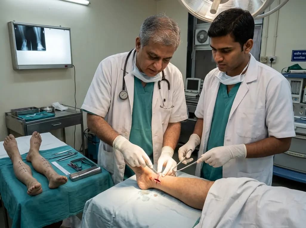
A paired cadaveric study compared triple and quintuple percutaneous Achilles tendon lengthening. While quintuple hemisection showed numerically greater elongation, differences were not statistically significant.
Today
A randomized trial in California evaluated a grocery support program for low-income, chronically ill adults. The intervention improved nutrition security by 2.8%, increased vegetable intake, boosted mental health scores, and reduced cost-related healthcare delays by 3.8%, showing the power of food-as-medicine.
Today
