.webp&w=384&q=75)

Loading, please wait...
Loading, please wait...

Accurately predicting meningioma recurrence risk remains a fundamental challenge in modern neurosurgery. For decades, neurosurgeons relied primarily on the classic Simpson grading system to estimate relapse likelihood. Donald Simpson established this surgical grading scale in 1957. It assesses the extent of macroscopic tumor resection and dural attachment removal. However, modern surgical environments have transformed significantly. Contemporary neurosurgeons now operate with advanced microsurgical techniques, ultrasonic aspirators, and intraoperative neuronavigation. Furthermore, high-resolution neuroimaging allows clinicians to inspect subtle anatomical margins with exceptional clarity. Because of these technical advancements, many researchers question whether subjective intraoperative impression still provides reliable prognostic guidance. Surgical line-of-sight limitations can obscure tiny fragments along critical venous sinuses or skull base crevices. Consequently, a surgeon may consider a resection complete, yet residual neoplastic cells remain behind. Therefore, clinicians urgently need objective radiographic metrics to refine postoperative risk stratification. Recent evidence highlights the crucial role of residual tumor volumetry on early postoperative imaging. By shifting focus from subjective surgeon impressions to quantitative MRI analysis, clinicians can better anticipate disease trajectories and optimize patient outcomes.
Intraoperative evaluations often diverge markedly from postoperative imaging findings. In a landmark retrospective study of 475 patients undergoing resection for intracranial WHO grade 1 to 3 meningiomas, investigators uncovered striking discordances. Specifically, surgeons designated gross total resection in 374 cases. However, postoperative magnetic resonance imaging demonstrated residual tumor in 7% of those patients. This discrepancy underscores how microscopic remnants easily evade direct visual inspection during complex cranial procedures. Even more remarkably, the inverse scenario occurred with notable frequency. In 13% of cases where the operating surgeon reported subtotal resection, postoperative imaging revealed no visible residual tumor tissue. Statistical analysis yielded a moderate Cohen’s kappa coefficient of 0.766, proving that subjective surgeon impressions and objective radiographic findings do not perfectly align. These observational differences carry profound clinical implications for ongoing patient surveillance. When neurosurgeons underestimate residual tissue, patients may miss necessary adjuvant therapies or follow-up scans. Conversely, overestimating residual burden might subject individuals to unwarranted anxiety or overtreatment. Therefore, postoperative magnetic resonance imaging serves as an indispensable corrective measure. It bridges the gap between surgical perception and radiographic reality, establishing an objective baseline for long-term monitoring.
Quantitative volumetric analysis provides deeper prognostic insight than traditional dichotomous resection categories. In univariate analyses, the extent of resection, absolute postoperative tumor volume, and relative volume reduction strongly correlated with tumor recurrence. Receiver operating characteristic curves confirmed that postoperative volume and relative volume reduction achieved comparable diagnostic accuracy, each yielding an area under the curve of 0.73. Interestingly, absolute volume reduction failed to demonstrate independent predictive utility. This finding indicates that tumor proportion removed matters significantly more than the raw mass debulked. In multivariate Cox regression modeling, only two variables retained independent predictive power for tumor recurrence: high-grade histopathology and relative volume reduction. High-grade histology increased recurrence hazard by more than fourfold. Concurrently, relative volume reduction significantly modulated long-term recurrence rates. Every percentage point of relative tumor debulking systematically reduced the hazard of relapse. Consequently, achieving maximal safe volumetric cytoreduction remains a vital therapeutic objective, even when complete surgical excision proves anatomically unachievable. These mathematical models demonstrate that volumetric metrics capture biological tumor behavior far more effectively than subjective categorical classifications. As a result, neuro-oncology teams should prioritize quantitative volumetric calculations when assessing recurrence risk and progression kinetics.
The integration of volumetric measurements directly informs multidisciplinary clinical decision-making. Historically, clinicians faced dilemmas when managing tumors encasing major dural venous sinuses, cranial nerves, or internal carotid branches. Aggressive attempts at radical Simpson grade 1 resections in these delicate anatomical locations often cause severe neurological morbidity. Fortunately, volumetric evidence confirms that achieving substantial relative volume reduction yields durable tumor control while safeguarding functional integrity. Surgeons can deliberately preserve critical neurovascular structures knowing that cytoreduction meaningfully dampens recurrence risk. Furthermore, exact volumetric assessment of residual disease empowers radiation oncologists. Modern stereotactic radiosurgery and fractionated stereotactic radiotherapy depend heavily on precise volumetric target delineation. When neurosurgeons obtain standardized postoperative volumetric MRI scans, radiation oncologists can contour target volumes with enhanced accuracy. Additionally, early volumetric monitoring helps identify residual lesions demonstrating aggressive growth trajectories before clinical symptoms manifest. Thus, multidisciplinary tumor boards can deploy adjuvant radiotherapy proactively rather than reactively. This strategic collaboration optimizes local disease control, minimizes radiation-induced toxicity, and preserves overall quality of life. Ultimately, volumetric metrics transform therapeutic paradigms from anatomical guesswork into data-driven precision oncology.
Establishing routine postoperative magnetic resonance imaging is essential for standardized clinical management. Although some health systems omit immediate postoperative imaging after perceived gross total resections, recent findings challenge this conservative policy. Omitting baseline imaging prevents clinicians from detecting occult residual lesions that lurk behind complex anatomical structures. Moreover, postoperative tissue distortion, surgical scar formation, and hemostatic materials complicate late imaging interpretation. Therefore, neurosurgeons should obtain contrast-enhanced magnetic resonance imaging within forty-eight to seventy-two hours following surgery. Early baseline imaging captures true residual tumor enhancement before reactive pachymeningeal enhancement develops. Additionally, establishing a precise volumetric baseline allows clinicians to differentiate indolent remnants from rapidly expanding recurrences during subsequent surveillance intervals. Health centers should incorporate automated or semi-automated volumetric segmentation software into routine Picture Archiving and Communication Systems. Such digital tools eliminate interobserver variability and deliver reproducible volumetric measurements across longitudinal clinical follow-up. In resource-conscious healthcare environments, this standardized approach prevents costly emergency interventions caused by undetected recurrences. Consequently, institutional imaging protocols must mandate early postoperative volumetry to safeguard surgical quality and guide long-term neuro-oncological care.
Postoperative magnetic resonance imaging detects unsuspected residual tumor in approximately seven percent of cases presumed completely resected intraoperatively. Early contrast-enhanced imaging establishes an objective baseline, rules out occult remnants along complex skull base crevices, and prevents misinterpretation of delayed reactive postoperative dural enhancement during long-term neuro-oncological surveillance.
Relative volume reduction measures the percentage of tumor excised relative to original burden. Multivariate models demonstrate that higher relative reduction independently reduces recurrence hazard. Maximizing volumetric cytoreduction significantly improves long-term disease control, confirming that aggressive safe debulking provides tangible clinical benefit even when total surgical excision remains technically infeasible.
The Simpson grading system relies heavily on subjective surgeon impressions and limited operative line of sight. Studies demonstrate frequent discordance with postoperative neuroimaging, including residual tumors after presumed gross total resection. Volumetric magnetic resonance imaging provides objective, reproducible quantitative measurements that predict tumor recurrence more reliably than classical surgical estimates.
Disclaimer: This content is for informational and educational purposes only... Refer to the latest local and national guidelines for clinical practice.
References

Read summarized clinical updates, watch expert medical content, and earn CME certifications right from your smartphone.
Open your smartphone camera to scan and install.





A retrospective analysis reveals that postoperative tumor volume and relative volume reduction on MRI outperform traditional Simpson grading in predicting meningioma recurrence risk, underscoring the vital clinical role of routine postoperative imaging.
Today

Survival analyses often report hazard ratios, but mathematical non-collapsibility creates crucial differences between conditional and marginal estimates. Clinicians must account for unmeasured confounding and model selection when interpreting trial endpoints and real-world cardiovascular outcomes.
Today

A cross-sectional study of 700 college students highlights significant contraceptive knowledge disparities between sexually active and inactive young adults. The findings demonstrate a critical need for proactive, comprehensive sexual health counseling before sexual debut.
Yesterday
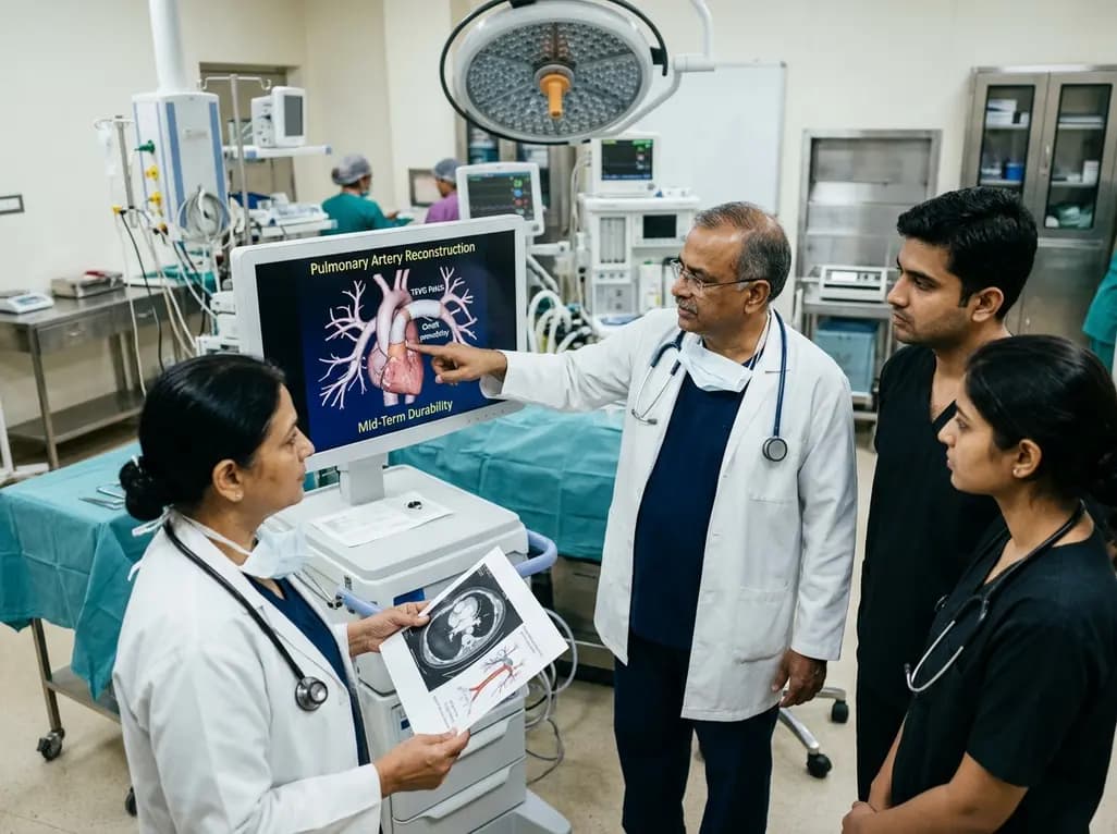
Autologous tissue-engineered vascular graft patches created via in-body tissue architecture show excellent mid-term durability and safety for pulmonary artery reconstruction in congenital heart surgery, particularly for peripheral branch augmentation.
Today

A novel cluster analysis of real-world datasets reveals four distinct CPPD disease phenotypes, validating existing EULAR categories while uncovering previously unrecognized monoarticular and axial presentations to refine patient management.
Today

Recent pharmacokinetic research demonstrates that trace dermal exposure to microgram quantities of trenbolone and metenolone produces detectable urinary metabolites for days to over a week, providing critical objective data for evaluating unintentional contamination claims in sports drug testing.
Today
