.webp&w=384&q=75)

Loading, please wait...
Loading, please wait...
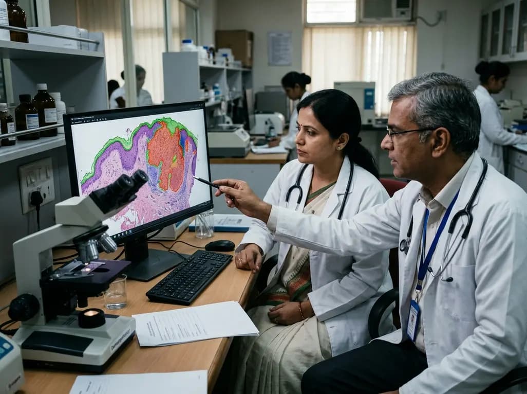
Accurate histopathological evaluation forms the cornerstone of clinical decision-making in cutaneous oncology. Pathologists routinely measure Breslow thickness and identify ulceration to determine pathological tumor stage. However, manual delineation of epidermis and tumor boundaries on whole-slide images remains time-consuming and prone to observer variation. Recent breakthroughs in computational pathology offer promising solutions to standardize diagnostic workflows. A primary prerequisite for clinical translation is robust algorithmic validation across curated datasets. In this context, researchers designed the Mel-DEPTHS dataset to accelerate reproducible advances in automated melanoma staging. This open benchmark addresses critical annotation challenges while establishing rigorous standards for semantic segmentation in digital dermatopathology.
Cutaneous melanoma represents one of the most aggressive skin malignancies globally. Consequently, precise risk stratification dictates surgical margins and systemic treatment pathways. Clinicians rely fundamentally on the American Joint Committee on Cancer staging system, where primary tumor depth directly influences survival prognosis. Pathologists calculate Breslow thickness by measuring the vertical distance from the granular layer of the epidermis to the deepest malignant cell. Additionally, the presence or absence of epidermal ulceration substantially alters tumor stage.
Despite its clinical significance, manual measurement faces substantial challenges in day-to-day practice. Histological specimens often exhibit architectural complexity, inflammatory infiltrates, and melanophages that confound precise boundary identification. Moreover, inter-observer variability across pathologists can lead to discordance in borderline measurements, potentially affecting stage allocation. Digitizing histology via whole-slide imaging provides an opportunity to apply computational tools for objective measurement. However, training deep neural networks requires massive quantities of pixel-level annotations. Generating manual segmentations across gigapixel images demands extensive time from specialized dermatopathologists. Therefore, the lack of standardized, publicly accessible datasets with expert ground truth has historically hindered the development of reliable artificial intelligence systems. Mel-DEPTHS directly addresses this critical barrier.
The Mel-DEPTHS dataset establishes a dedicated resource designed to standardize digital pathology research. Specifically, the cohort comprises fifty anonymized whole-slide images of cutaneous melanoma scanned at forty-times magnification with high optical resolution. Each digital slide features exhaustive, pixel-level masks that delineate both the overlying epidermis and the invasive tumor compartments. Crucially, the dataset includes paired clinical variables such as microscopic invasion depth, ulceration status, and verified pathological tumor stage.
To promote sound scientific evaluation, the authors established fixed training and testing partitions. This structural standardization eliminates data leakage and enables direct comparisons across future computational studies. Furthermore, the dataset captures diverse morphological subtypes, varying levels of solar elastosis, and distinct architectural growth patterns. Consequently, models trained on this benchmark must generalize across wide histological variability rather than overfitting to uniform tissue sections. By delivering expert-validated ground truth alongside clinical parameters, Mel-DEPTHS bridges the gap between pure computer vision metrics and actionable diagnostic variables. Pathologists and computer scientists can utilize this benchmark to validate models that quantify anatomical features directly tied to patient staging and surgical planning.
Pixel-level semantic segmentation on whole-slide images represents a formidable operational bottleneck due to immense image dimensions. To mitigate this annotation burden, the study introduced the Expert-Supervised Iterative Self-Training framework. This protocol systematically integrates algorithmic efficiency with dermatopathologist oversight to generate high-fidelity ground truth masks.
Initially, a baseline segmentation network generates preliminary pseudo-labels across unannotated histological slides. Subsequently, board-certified dermatopathologists review these automated boundaries, correcting errors in challenging regions such as poorly differentiated tumor nests and fragmented epidermis. The computational team then retrains the segmentation algorithms using these refined masks. Consequently, the updated model generates superior pseudo-labels for subsequent iterations. This human-in-the-loop paradigm drastically reduces manual contouring time while preserving strict histological fidelity. Furthermore, iterative refinement minimizes individual reader bias, establishing consistent contour definitions across the entire dataset. Ultimately, the framework demonstrates how structured clinical oversight can accelerate large-scale dataset creation without sacrificing analytical rigor or diagnostic validity.
To establish performance baselines on Mel-DEPTHS, investigators evaluated six state-of-the-art segmentation architectures. The benchmark compared classical convolutional models, including UNet, UNet++, and UNet3+, against contemporary frameworks such as UPerNet, ConvUNeXt, and TransUNet. Evaluation metrics spanned whole-slide precision, recall, Intersection over Union, and Dice similarity coefficients.
Across comprehensive testing, hybrid transformer-based models demonstrated clear analytical superiority. TransUNet achieved the highest overall segmentation accuracy, closely followed by ConvUNeXt and UPerNet. Specifically, TransUNet excelled in capturing long-range contextual relationships while preserving fine edge resolution along epidermal basements. Three-fold cross-validation confirmed consistent ranking among architectures, verifying the robustness of the expert annotations. While conventional convolutional networks struggled with irregular tumor margins, vision transformer backbones effectively resolved complex tissue transitions. In addition, the models maintained robust performance across slides with dense lymphocytic infiltrates. These benchmark results provide crucial guidance for engineering production-grade diagnostic tools in digital oncology.
Translating segmentation algorithms into routine pathology workflows requires stringent validation beyond aggregate statistical metrics. Clinicians must trust that automated boundaries accurately reflect true tissue architecture. Mel-DEPTHS supports this translation by aligning segmentation outputs with prognostic markers like Breslow thickness and epidermal ulceration.
When algorithms delineate epidermal margins and tumor borders accurately, software can compute vertical invasion depth automatically. Consequently, automated measurements can decrease measurement variability and reduce turnaround times in busy pathology laboratories. Furthermore, objective ulceration detection eliminates subjective ambiguity in assessing denuded epithelial layers. Pathologists can review model overlays as an assistive diagnostic aid, ensuring rapid verification rather than manual re-measurement. Additionally, standardized benchmarks encourage algorithm developers to address real-world artifacts, including tissue folds, out-of-focus fields, and staining inconsistencies. By demonstrating reliable segmentation across varied slides, the dataset provides a solid foundation for regulatory clearance and prospective clinical trials in computational dermatopathology.
The release of Mel-DEPTHS marks an important milestone toward automated diagnostic assistance in skin cancer care. Nevertheless, ongoing expansion remains essential to accommodate broader histological diversity. Future efforts will likely expand the dataset to include rare melanoma variants, special anatomical sites, and multi-institutional scanning platforms.
Moreover, integrating segmentation algorithms with multimodal clinical data will further enhance personalized oncology. Combining objective tumor depth with genomic profiles, sentinel lymph node status, and dermoscopic imaging can refine risk stratification models. Clinicians could leverage these combined insights to optimize adjuvant immunotherapy selection and surgical margin planning. As computational pathology platforms mature, benchmark datasets like Mel-DEPTHS will remain foundational for rigorous quality assurance. Ultimately, fostering collaborative development between dermatopathologists and artificial intelligence engineers ensures that computational tools enhance diagnostic accuracy and elevate patient care standards worldwide.
Mel-DEPTHS provides high-resolution, pixel-level masks for both the epidermis and invasive tumor compartments across whole-slide images. Consequently, computational algorithms can identify the epidermal granular layer and the deepest invading melanoma cells with sub-micron precision. This automation standardizes vertical invasion depth calculations, directly reducing the inter-observer variability frequently encountered during manual ocular micrometer assessment and improving clinical staging accuracy.
The Expert-Supervised Iterative Self-Training protocol overcomes the immense operational burden of manual whole-slide annotation. Initially, a baseline algorithm generates preliminary pseudo-labels across unannotated tissue slides. Expert dermatopathologists then review and correct these automated boundaries. Retraining models on these refined masks progressively enhances segmentation quality. This structured human-in-the-loop workflow ensures gold-standard annotation fidelity while substantially accelerating large-scale dataset curation.
TransUNet combines convolutional feature extraction with self-attention vision transformers, allowing the model to capture global contextual dependencies across gigapixel tissue sections. Meanwhile, its decoder retains high-resolution local spatial details along tissue margins. This hybrid design allows TransUNet to distinguish subtle tumor boundaries and fragmented epidermal layers far more effectively than traditional convolutional networks, resulting in superior Dice similarity scores.
Disclaimer: This content is for informational and educational purposes only and does not constitute medical advice, diagnosis, or treatment. Healthcare professionals should exercise independent clinical judgment and consult relevant clinical guidelines, drug prescribing information, and institutional protocols before making diagnostic or treatment decisions. Patients should consult a qualified physician regarding any medical conditions. Refer to the latest local and national guidelines for clinical practice.
References

Read summarized clinical updates, watch expert medical content, and earn CME certifications right from your smartphone.
Open your smartphone camera to scan and install.





Mel-DEPTHS is a standardized benchmark dataset offering pixel-level epidermis and tumor segmentation on whole-slide images. By combining expert annotations with deep learning models like TransUNet, this dataset enhances automated melanoma staging, Breslow thickness measurement, and histopathological reproducibility.
last month
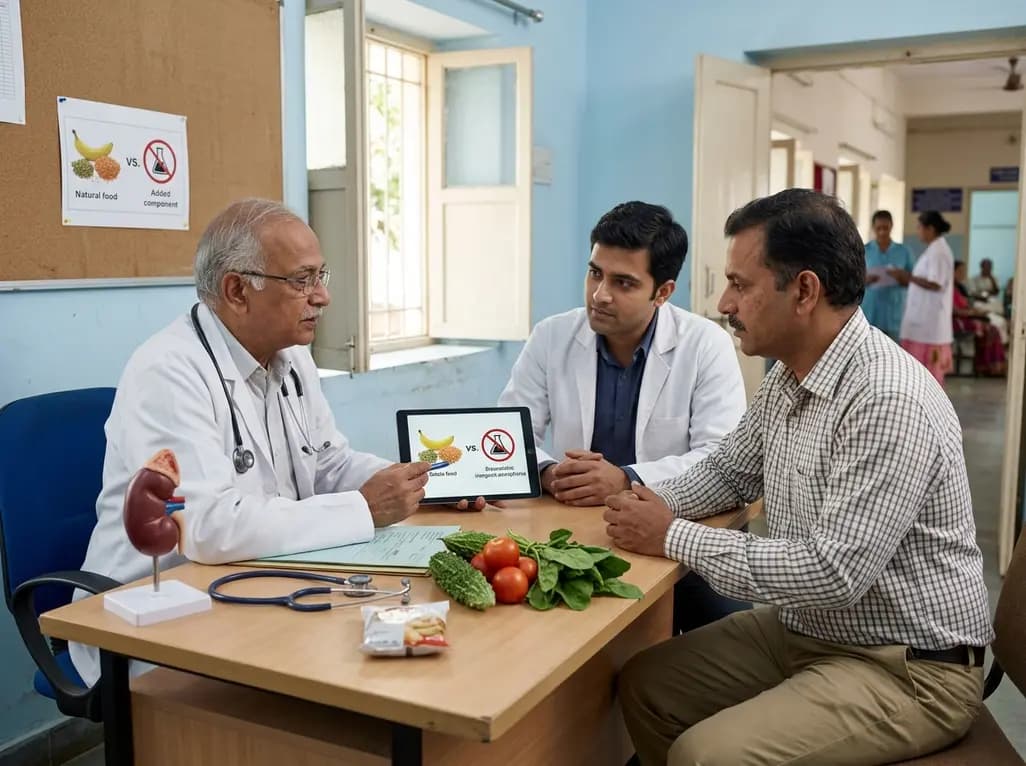
Modern renal nutrition is shifting toward additive-aware dietary guidance. This paradigm differentiates between intrinsic nutrients in whole foods and hyper-bioavailable inorganic additives, offering practical strategies to mitigate additive stacking and toxicity without restricting nutritious whole foods.
Today

A multiparametric MRI approach combining quantitative T2 shading, apparent diffusion coefficient (ADC) measurements, and morphological signs provides superior diagnostic accuracy over subjective evaluation to differentiate ovarian endometriomas from benign hemorrhagic cysts.
Today

A landmark case report reveals a donor heart beating for a century in a 63-year-old recipient, challenging conventional age limits in cardiac transplantation and offering critical lessons on organ longevity, donor selection, and long-term graft survival.
Today
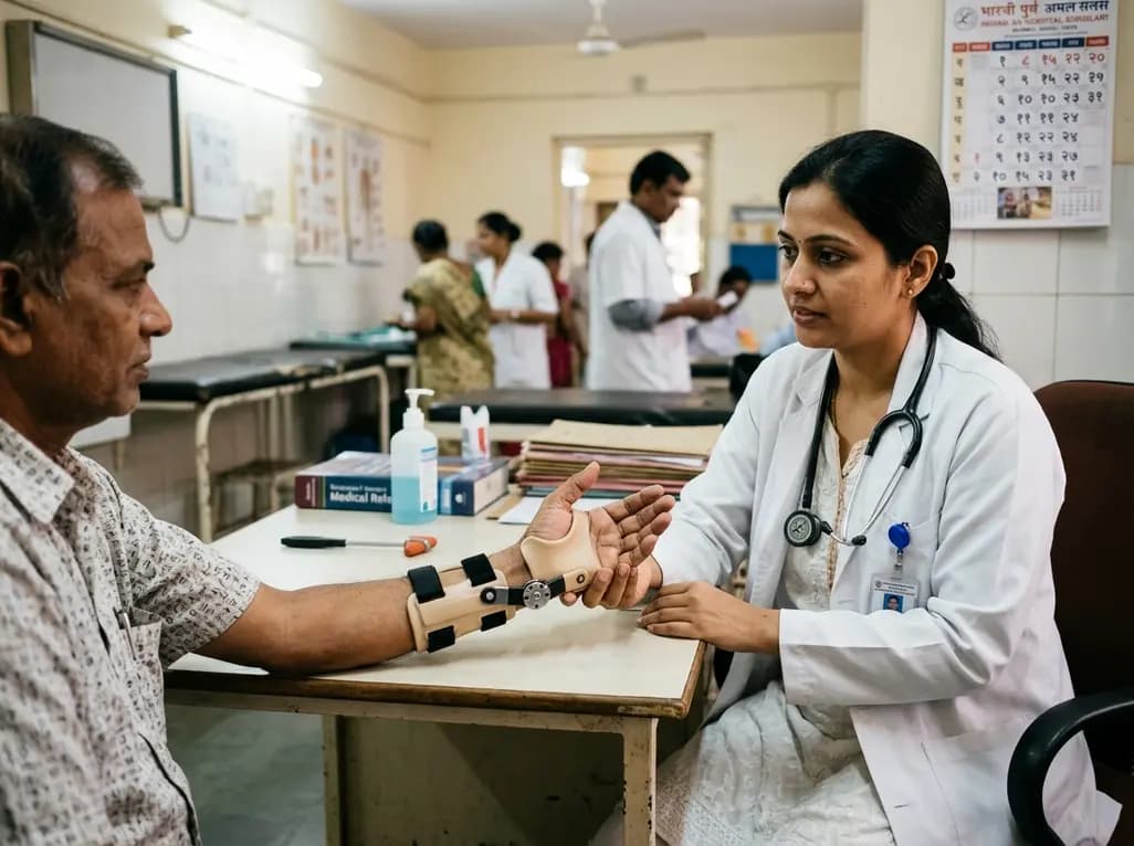
A new randomized crossover trial reveals that a hinged ulnar gutter orthosis significantly improves hand function and weight-bearing tolerance compared to standard volar splints in patients with degenerative triangular fibrocartilage complex lesions, supporting functionally permissive orthotic designs.
Today

New research reveals that M1 macrophage-derived exosomes aggravate diabetic nephropathy by transferring WTAP to stabilize S1PR2 mRNA. Silencing WTAP in these vesicles attenuates endothelial injury and renal fibrosis, pointing toward innovative nanomedicine therapies.
Yesterday
