.webp&w=384&q=75)

Loading, please wait...
Loading, please wait...
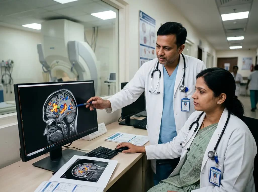
Accurate mapping of cortical language networks is essential for advancing basic cognitive neuroscience and guiding safe neurosurgical resections. Recent advances demonstrate that MEG language mapping using individual functional localizers significantly improves diagnostic precision over traditional group-averaging approaches. By isolating language-responsive sensors within each patient, neuroscientists and clinicians can capture critical temporal dynamics while avoiding the anatomical blurring that often complicates clinical interventions.
Preoperative localization of eloquent cortex remains a critical component of modern neurosurgical planning, especially for patients with focal epilepsy or peri-Sylvian tumors. Traditional functional mapping approaches often rely on functional magnetic resonance imaging or invasive cortical stimulation. However, magnetoencephalography offers a distinct advantage because it directly records postsynaptic neuronal currents with millisecond-level temporal resolution. This high temporal precision enables clinicians to track the rapid, sequential stages of phonological, lexical, and syntactic decoding in real time.
Furthermore, cortical language organization varies markedly among individuals, both anatomically and functionally. In standard clinical practice, relying on rigid anatomical landmarks can introduce substantial errors, because language areas reside in close proximity to domain-general cognitive networks. Consequently, MEG language mapping provides clinicians with a non-invasive, physiologically direct tool to characterize hemispheric dominance and delineate critical boundaries. By capturing both spatial topographies and oscillatory electrophysiological signatures, clinicians can identify eloquent cortical units with exceptional reliability. Therefore, adopting subject-specific MEG paradigms represents a vital step toward personalizing presurgical assessments, minimizing postoperative neurological deficits, and preserving crucial communicative faculties in neurosurgical candidates.
For decades, cognitive neuroimaging paradigms have relied on transforming individual brain scans into standardized stereotactic spaces, such as Talairach or MNI templates. Although group-level averaging enhances statistical power across large participant cohorts, it creates major disadvantages when evaluating the language network. Structural morphology within the lateral frontal and superior temporal cortices demonstrates remarkable inter-individual variability. Consequently, spatial normalization often misaligns functional areas across subjects, smoothing out critical signal boundaries and suppressing individualized neural signatures.
Moreover, group averaging tends to mask subtle functional variations that occur naturally across healthy individuals and clinical populations. When brain scans are merged indiscriminately, weak or spatially idiosyncratic activations can disappear entirely, producing misleading maps of eloquent function. In neurosurgical and neurological practice, an averaged group template cannot account for the patient-specific anatomical distortions caused by mass effect, edema, or long-standing neuroplastic reorganization. Therefore, clinicians who depend on group-level assumptions risk mischaracterizing essential cortical hubs. To overcome these inherent constraints, neuroimaging researchers must transition toward subject-specific mapping methodologies that honor individual anatomical nuances while retaining robust statistical power.
To resolve inter-individual variability, researchers have successfully adapted functional localizer paradigms from visual neuroscience and fMRI into magnetoencephalography protocols. In these investigations, participants undergo targeted contrast tasks—such as processing natural sentences compared to structurally matched nonword sequences—to isolate specific language-sensitive responses. Recent experimental evidence across multiple languages, including Dutch and English cohorts, demonstrates that sensor and source topographies of language responses remain remarkably stable within an individual over time.
However, these identical functional topographies display substantial spatial variation across different participants. Consequently, analyzing MEG data at the individual sensor level reveals language-responsive neural units that traditional group-averaging methods systematically overlook. This subject-specific strategy dramatically increases signal detection sensitivity, allowing investigators to isolate eloquent temporal and frontal regions with greater functional fidelity. Furthermore, individual functional localization preserves fine-grained electrophysiological dynamics, such as evoked field peaks and oscillatory power changes in the beta and gamma bands. As a result, this refined methodology provides an objective, reproducible framework for characterizing language architecture across diverse clinical and cognitive research cohorts.
For neurosurgeons and clinical epileptologists, the clinical translation of individualized MEG localizers represents a significant diagnostic advancement. Presurgical evaluation requires absolute precision to maximize lesion resection while sparing critical eloquent cortex. Historically, the intracarotid amobarbital procedure (Wada test) served as the gold standard for language lateralization, despite carrying inherent procedural risks such as stroke, arterial dissection, and prolonged hospital stays. Non-invasive MEG mapping offers a safer, highly concordant alternative that determines hemispheric dominance without invasive catheterization.
Additionally, MEG localizer protocols provide exquisite spatial coordinates for intraoperative guidance and neuronavigation systems. By accurately demarcating receptive and expressive language hubs prior to surgery, surgical teams can tailor craniotomy margins and optimize the placement of subdural grid electrodes for direct cortical stimulation. Moreover, in pediatric patients or individuals with pharmacoresistant epilepsy who cannot tolerate lengthy awake craniotomies, passive or task-based MEG mapping delivers indispensable functional insights. Consequently, integrating individualized MEG protocols into the presurgical pathway optimizes operative trajectories, reduces operative time, and significantly lowers the likelihood of permanent postoperative aphasia.
Looking forward, the true strength of individualized MEG language mapping lies in its integration within multimodal neuroimaging pipelines. While functional MRI provides outstanding spatial resolution, it relies on hemodynamic changes that lag behind rapid neural activity by several seconds. In contrast, MEG directly captures electrophysiological currents on a millisecond scale. Therefore, combining high-resolution structural MRI, diffusion tractography, and individualized MEG creates a comprehensive structural and functional roadmap of language white-matter pathways and cortical hubs.
Furthermore, emerging technological innovations, such as optically pumped magnetometers (OPMs), are revolutionizing magnetoencephalography. Wearable OPM sensors eliminate the need for rigid cryogenic dewars, permitting unconstrained head movement and making scanning accessible for young children and claustrophobic patients. In parallel, advanced machine-learning algorithms and beamforming source-reconstruction techniques continue to enhance signal-to-noise ratios in individualized data sets. Consequently, these technical refinements will solidify the clinical utility of MEG across major neurological centers worldwide. Ultimately, embracing subject-specific electrophysiological mapping will accelerate our understanding of neuroplasticity, stroke recovery, and complex cognitive processing in both clinical and academic domains.
MEG language mapping measures direct neuronal magnetic flux with millisecond temporal precision, whereas functional MRI measures indirect blood-oxygen-level-dependent hemodynamic changes occurring over several seconds. While fMRI provides superior spatial resolution, MEG captures real-time electrophysiological dynamics and avoids neurovascular uncoupling errors caused by brain tumors or vascular malformations. Therefore, utilizing both modalities concurrently provides complementary spatial and temporal insights that maximize presurgical accuracy and patient safety.
Individual functional localization accounts for the high anatomical and functional variability naturally present in lateral frontal and temporal language cortices. Group-averaging methods project individual brains onto a common template, which inadvertently blurs distinct cortical borders and dilutes signal sensitivity. In contrast, individualized localizers precisely isolate language-responsive sensors within each subject, ensuring that unique functional hubs and pathological structural shifts are accurately identified without loss of spatial specificity.
Extensive clinical research demonstrates that MEG language mapping exhibits high concordance with the Wada test in determining hemispheric language dominance. Because MEG is completely non-invasive, it eliminates procedural risks such as arterial dissection, contrast reactions, and transient ischemic events. Consequently, many comprehensive epilepsy centers now utilize MEG as a primary non-invasive lateralization tool, reserving invasive amobarbital testing exclusively for highly complex or ambiguous clinical cases.
Disclaimer: This content is for informational and educational purposes only. It is not intended to be a substitute for professional medical advice, diagnosis, or treatment. Always seek the advice of your physician or other qualified health provider with any questions you may have regarding a medical condition. Never disregard professional medical advice or delay in seeking it because of something you have read on this website. Refer to the latest local and national guidelines for clinical practice.
References

Read summarized clinical updates, watch expert medical content, and earn CME certifications right from your smartphone.
Open your smartphone camera to scan and install.





Individualized MEG language mapping overcomes inter-individual anatomical variability by identifying language-responsive sensors within individual brains, significantly boosting sensitivity and functional resolution for clinical neuroscience and presurgical planning.
last month

A nationwide multicenter study identified three COPD clinical phenotypes, finding that mild smoking males experience the highest rate of rapid lung function decline. A CatBoost model using six routine clinical and socioeconomic variables accurately stratifies high-risk patients for early therapeutic intervention.
Today
Discover the crucial distinctions between mesonephric and mesonephric-like proliferations of the female genital tract. Learn the key histomorphologic, immunohistochemical, and molecular differences essential for accurate diagnosis and clinical management.
Today

A large UK Biobank study reveals that plasma proteomic signatures capture preclinical organ damage and improve multiorgan risk prediction across early CKM syndrome stages 0 to 2 beyond traditional PREVENT clinical models.
Today
A narrative review investigates whether the articularis genus muscle functions as an independent anatomical entity or blends with the vastus intermedius. We evaluate its morphology, role in retracting the suprapatellar bursa, and direct clinical significance in anterior knee pain and arthroplasty.
Today

A Bayesian multilevel meta-analysis reveals that aerobic training combined with moderate carbohydrate restriction modestly lowers HbA1c in type 2 diabetes. However, sparse data and very low certainty leave incremental benefits over exercise or diet alone unproven, highlighting the need for individualized care.
Today
