.webp&w=384&q=75)

Loading, please wait...
Loading, please wait...
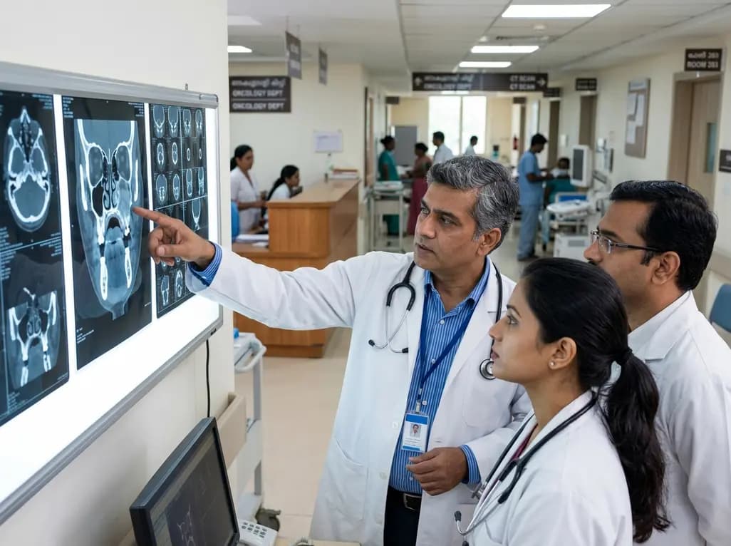
Upper gingiva and hard palate squamous cell carcinoma presents unique therapeutic challenges due to nearby facial anatomy. Specifically, maxillary sinus mucosal extension serves as a critical radiographic indicator of aggressive vertical tumor infiltration. Historically, oncologists debated the prognostic value of antral mucosal involvement because concurrent bone invasion frequently obscured independent clinical risks. However, recent evidence confirms that radiological mucosal extension independently correlates with poorer overall survival and disease-specific survival. In routine head and neck oncology, early identification of this vertical trajectory directly impacts clinical decision-making. Tumors of the upper gingiva lie near the thin cortical floor of the maxillary sinus. Consequently, malignant cells traverse the bony barrier to reach the schneiderian mucosa. This anatomical progression transforms an oral malignancy into a complex sinonasal disease process. Surgeons and radiologists must therefore evaluate mucosal thickening within the maxillary antrum with meticulous precision. Accurately distinguishing true neoplastic mucosal extension from reactive inflammatory sinusitis remains essential for proper staging. When multidisciplinary oncology teams recognize this radiographic feature early, they can plan definitive resections that avoid microscopic residual disease.
Modern management of upper jaw malignancies relies heavily on multimodality cross-sectional diagnostic imaging. High-resolution computed tomography provides detailed bone window views that accurately delineate cortical erosion and osteolytic destruction. Conversely, magnetic resonance imaging excels at evaluating soft-tissue contrast, allowing clinicians to distinguish neoplastic infiltration from retained inflammatory secretions. Preoperative computed tomography and magnetic resonance imaging together illuminate the vertical trajectory of upper gingival carcinomas. However, interpreting antral mucosal changes remains challenging because secondary sinusitis frequently accompanies tumor obstruction of the ostiomeatal complex. Radiologists look for specific radiological indicators, such as asymmetric mucosal enhancement, nodular thickening, and frank soft-tissue mass continuity. These cross-sectional features confirm true neoplastic extension into the sinus cavity rather than reactive edematous mucosa. Furthermore, contrast-enhanced sequences help identify perineural spread along palatine and infraorbital nerve branches. Clinicians must actively correlate these imaging characteristics with clinical findings during staging examinations. Accurately characterizing mucosal invasion ensures precise assignment within the tumor staging framework. When imaging underrepresents tumor vertical depth, surgeons risk understaging advanced lesions. As a result, comprehensive multiplanar radiological evaluation directly dictates whether an operation requires conservative partial resection or extended radical maxillectomy.
Clinical researchers have long questioned whether mucosal invasion independently impacts prognosis or simply reflects aggressive cortical bone erosion. To address this confounding issue, investigators conducted detailed subgroup analyses isolated strictly to patients with confirmed radiological bone invasion. The statistical findings revealed that disease-specific survival remained significantly worse among patients exhibiting radiological mucosal extension compared to those without antral involvement. Therefore, mucosal involvement represents an adverse biological marker beyond mere osseous destruction. Overall survival rates also showed substantial reductions when mucosal invasion was present on preoperative scans. Patients lacking antral mucosal spread maintained superior local control rates, translating directly into durable disease remission. Conversely, individuals demonstrating vertical mucosal extension experienced elevated rates of locoregional recurrence and shortened survival intervals. These observations demonstrate that mucosal involvement denotes an aggressive, infiltrating phenotype capable of rapid microscopic advancement. In addition, the persistent survival gap observed in T4 cohorts confirms that osseous destruction alone does not explain patient mortality. Clinicians must consequently recognize that antral mucosal infiltration signals a high-risk oncologic subtype. Treating physicians should utilize this critical radiological marker during multidisciplinary tumor board discussions to stratify patient prognosis accurately.
Achieving microscopically negative resection margins remains the cornerstone of curative oncologic surgery for upper gingival and hard palate carcinomas. However, multivariable statistical models demonstrate that adjusting for surgical margin status significantly attenuates the prognostic impact of sinus mucosal extension. This critical finding underscores that incomplete tumor clearance, rather than mucosal infiltration alone, directly drives treatment failure. When tumors extend into the maxillary antrum, surgical extirpation becomes exponentially more complex. Surgeons frequently encounter tight three-dimensional spaces, friable respiratory mucosa, and obscured anatomical planes. Consequently, achieving wide, tumor-free margins in the upper maxilla poses considerable technical difficulty. Positive or close margins allow microscopic residual cancer cells to proliferate rapidly within the sinonasal cavities. Thus, the adverse prognosis historically linked with antral mucosal extension stems largely from positive surgical margins. Surgical teams must therefore anticipate mucosal involvement prior to entering the operating room. Surgeons can design wider resections, such as subtotal or infrastructure maxillectomies, rather than attempting marginal bone resections. Furthermore, intraoperative frozen-section margin analysis provides essential guidance. When oncologists secure clear pathological margins, patient survival rates improve markedly despite deep vertical tumor penetration.
Oral cancers represent an immense public health burden across India, primarily driven by widespread smokeless tobacco, gutka, and betel quid usage. Squamous cell carcinoma of the upper alveolar ridge and palate often manifests at advanced stages because patients delay clinical consultation. Consequently, Indian head and neck oncologists routinely treat tumors presenting with substantial palatal destruction and maxillary sinus infiltration. Managing these challenging cases demands close coordination among surgical oncologists, radiation oncologists, maxillofacial prosthodontists, and reconstructive surgeons. When imaging demonstrates sinus mucosal involvement, clinicians must consider aggressive multimodal management rather than standalone conservative surgery. Adjuvant radiation therapy or concurrent chemoradiotherapy becomes indispensable whenever surgical margins remain narrow or microscopically positive. Additionally, resecting upper maxillary lesions creates significant oronasal communication that severely impairs speech, mastication, and deglutition. Surgical teams must therefore plan definitive reconstruction concurrently. Options include vascularized free flaps, such as anterolateral thigh or fibula flaps, or customized dental obturators. Tertiary cancer centers throughout India benefit greatly from incorporating standardized preoperative sinus evaluations into multidisciplinary tumor boards. By identifying antral mucosal involvement early, oncologists can optimize surgical margins, select appropriate adjuvant protocols, and restore functional quality of life.
Radiological sinus mucosal extension occurs when cross-sectional imaging reveals abnormal mucosal thickening or nodular soft-tissue enhancement within the maxillary sinus directly adjacent to an invasive oral tumor. Computed tomography delineates the underlying bony erosion along the sinus floor, while magnetic resonance imaging reliably distinguishes neoplastic mucosal infiltration from retained secretions. Clinicians confirm true tumor extension by identifying continuous soft-tissue enhancement extending across the disrupted cortical floor into the sinus cavity.
Sinus mucosal extension significantly impairs disease-specific survival because vertical tumor penetration complicates surgical clearance within complex three-dimensional anatomical boundaries. When cancer infiltrates the schneiderian mucosa, malignant cells spread rapidly along the mucosal lining, increasing the incidence of positive or close surgical resection margins. This incomplete local clearance dramatically elevates the risk of persistent locoregional recurrence. Consequently, patients exhibiting antral mucosal spread experience substantially worse oncologic outcomes and shorter overall survival.
When preoperative scans demonstrate sinus mucosal involvement, surgeons should avoid conservative marginal alveolar resections. Instead, operative teams must execute planned subtotal or infrastructure maxillectomies to achieve adequate microscopic clearance beyond the sinus mucosa. Intraoperative frozen-section pathological evaluation of antral margins provides indispensable real-time guidance during resection. Furthermore, surgical oncologists should collaborate closely with reconstructive surgeons to immediately repair the resulting palatomaxillary defect using microvascular free flaps or custom obturators.
Disclaimer: This content is for informational and educational purposes only... Refer to the latest local and national guidelines for clinical practice.
References
1. Itotagawa M et al. Radiological maxillary sinus mucosal extension is associated with prognosis in upper gingiva and hard palate squamous cell carcinoma. Int J Oral Maxillofac Surg. 2026 Sep 12. doi: undefined. PMID: 42731955.
2. Nagasaki University Hospital Study Group. Prognostic Factors in Squamous Cell Carcinoma of the Maxillary Gingiva and Hard Palate: A Retrospective Analysis. PMC. 2025.
3. Shibuya H, et al. Squamous cell carcinoma of the maxillary sinus and the oral part of the upper jaw. Acta Oncol. 1994;33(1):43-47.

Read summarized clinical updates, watch expert medical content, and earn CME certifications right from your smartphone.
Open your smartphone camera to scan and install.





Radiological maxillary sinus mucosal extension serves as a vital prognostic marker in upper gingiva and hard palate squamous cell carcinoma. While associated with poorer survival even in bone-invasive tumors, its prognostic impact is largely mediated by surgical margin status, emphasizing radical clearance.
Today

At the New Delhi Summit, BRICS leaders adopted a landmark declaration committing member nations to united action against tuberculosis, non-communicable diseases, and future pandemic threats. Key actions include NIMHANS-led mental health networks, digital health architectures, and evidence-based integrative medicine.
Today
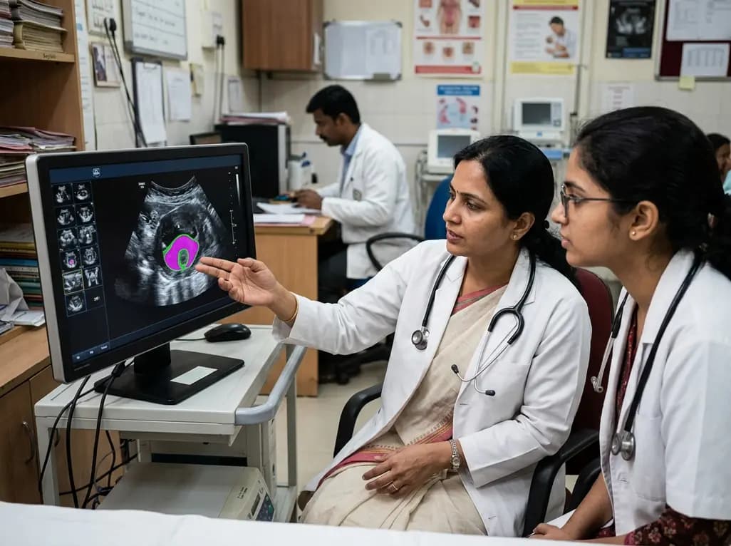
New evidence evaluates MoUNets, a static-weighted ensemble of UNet experts for automated levator hiatus segmentation in pelvic floor ultrasound. Learn how deep learning architectures perform in pelvic organ prolapse diagnostics and clinical practice.
Today

Mitochondrial deubiquitinases regulate organelle turnover by editing ubiquitin tags on damaged mitochondria. Discover how targeting these enzymes, notably USP30, offers therapeutic strategies for neurodegenerative, cardiovascular, and metabolic disorders.
Today

A rare case of a pediatric pathological femoral fracture caused by occult telangiectatic osteosarcoma demonstrates the risks of misdiagnosis after trauma, where intramedullary nailing prompted iatrogenic tumor dissemination and required modular total endoprosthetic reconstruction.
Yesterday

A cross-sectional study reveals that elevated METS-IR is independently associated with higher myopia odds in adolescents, showing biological synergy with obesity and highlighting the need for metabolic screening in pediatric eye care.
Today
