.webp&w=384&q=75)

Loading, please wait...
Loading, please wait...

Gestational nutritional health plays a defining role in shaping fetal organogenesis and long-term neural maturation. Recent neuroimaging data confirm that maternal anaemia significantly impedes infant neurodevelopment, altering cerebral architecture within the first two years of life. Historically, clinicians viewed mild reductions in maternal hemoglobin as a tolerable physiological adaptation. However, emerging structural evidence demonstrates that even modest maternal haematological compromises exert measurable detriments on infant neuroanatomy.
Researchers from King's College London and the University of Cape Town recently published groundbreaking neuroimaging findings. Specifically, their study evaluated mother-infant dyads to explore how gestational haematological deficits influence early brain maturation. Notably, the investigators deployed innovative ultra-low-field portable magnetic resonance imaging scanners alongside traditional high-field systems. This portable neuroimaging equipment allowed clinicians to scan vulnerable infants repeatedly in community settings between three and twenty-four months of age. Consequently, the team gathered objective morphological metrics across pivotal developmental windows.
The resulting dataset revealed that infants exposed to maternal anaemia exhibited 3.77 percent smaller total intracranial volumes. Furthermore, these structural discrepancies became clearly detectable around twelve months of age and persisted across subsequent evaluations. Most mothers in the South African cohort experienced only mild anaemia during gestation. Nevertheless, their offspring demonstrated substantial and enduring reductions in overall cerebral volume. In addition, these morphological changes highlight the critical necessity of proactive antenatal surveillance. Therefore, clinical teams must appreciate that even modest gestational haematological compromises significantly disrupt early infant brain architecture.
The Cape Town investigation followed 394 mother-infant pairs through rigorous longitudinal assessments. Within this cohort, researchers conducted serial neuroimaging scans on a designated infant subgroup at multiple developmental milestones. Importantly, the analysis revealed regional brain vulnerabilities that extended beyond global intracranial volume reductions. The researchers identified prominent deficits in three distinct anatomical structures: the putamen, the caudate nucleus, and the corpus callosum. In fact, infants exposed to intrauterine anaemia exhibited a 3.32 percent putamen volume reduction across two years.
Moreover, the researchers documented a striking temporal divergence in white matter expansion. At twelve months of age, infants of anaemic mothers displayed a 4 percent smaller corpus callosum compared to controls. However, by twenty-four months of age, this anatomical deficit widened dramatically to 6 percent. Thus, longitudinal observation demonstrates that the gap between healthy infants and anaemia-exposed peers expands during early life. In addition, these neuroimaging findings corroborate earlier observations in older cohorts that demonstrated lasting morphological deficits. Ultimately, these data confirm that gestational anaemia produces lasting structural footprints that compound over early childhood development.
The specific brain structures affected by maternal haematological deficits play pivotal roles in complex cognitive and motor functions. Specifically, the putamen and caudate nucleus constitute key components of the basal ganglia circuitry. These deep subcortical nuclei coordinate voluntary motor control, procedural memory, reinforcement learning, and executive functioning. Furthermore, the striatal circuits modulate emotional processing and impulse control through dense connections with the prefrontal cortex. When anaemia compromises early striatal development, children face heightened risks of attentional deficits, fine motor delays, and emotional dysregulation.
Similarly, the corpus callosum represents the principal white matter commissure connecting the cerebral hemispheres. This massive axonal tract facilitates interhemispheric communication, sensory integration, and bilateral motor coordination. Because the corpus callosum undergoes rapid myelination during the first two postnatal years, it requires substantial metabolic resources. Consequently, maternal iron deficiency deprives developing oligodendrocytes of critical cofactors necessary for timely myelin sheath formation. Hence, the observed 6 percent reduction in callosal volume may impair complex cross-cortical processing speed. Therefore, pediatricians must recognize that early anatomical compromises may eventually manifest as learning and behavioural obstacles at school age.
Gestational anaemia disrupts neurodevelopment through interconnected biochemical and cellular pathways. Iron serves as an indispensable micronutrient for mitochondrial electron transport, oxidative phosphorylation, and cellular energy synthesis. During gestation, the fetal central nervous system exhibits an exceptionally high metabolic demand to fuel rapid neurogenesis and synaptogenesis. Furthermore, neural progenitor cells rely heavily on iron-dependent enzymes for DNA synthesis and cellular proliferation. When maternal red cell mass drops, the placenta attempts to prioritize fetal iron transport through transferrin receptor upregulation. However, severe maternal depletion overwhelms these compensatory mechanisms, leading to restricted fetal iron accretion.
Consequently, the developing brain experiences subtle histochemical alterations long before clinical haematological signs manifest in the neonate. Specifically, iron deficiency curtails the enzymatic activity of ribonucleotide reductase and cytochromes within metabolically active deep nuclei. In addition, oligodendrocytes demonstrate exquisite sensitivity to iron restriction because they require iron for myelin lipid biosynthesis. As a result, hypomyelination occurs alongside diminished dendritic arborization across central neural pathways. Moreover, fetal hypoxia resulting from reduced maternal oxygen delivery compounds cellular oxidative stress. Therefore, these combined pathophysiological insults permanently alter microstructural neural connectivity and restrict macroscopic volumetric growth.
These neuroimaging findings carry immense clinical significance for healthcare providers practicing across India. According to recent National Family Health Survey estimates, more than fifty percent of pregnant Indian women suffer from anaemia. Historically, clinicians have focused primarily on acute obstetric risks, such as postpartum haemorrhage, preterm birth, and low birth weight. However, this new neuroimaging evidence proves that maternal haematological status actively governs the child's neurological future. In low- and middle-income settings, maternal anaemia frequently coexists with malnutrition, intestinal helminths, and systemic inflammation. Therefore, obstetricians and general practitioners must adopt aggressive, multifaceted strategies to correct iron deficiency before conception and throughout pregnancy.
Specifically, healthcare teams must implement universal early screening utilizing complete blood counts and serum ferritin testing. Furthermore, clinicians should enforce strict compliance with national guidelines under the Anemia Mukt Bharat initiative. Daily prophylactic elemental iron and folic acid supplementation remains essential for non-anaemic mothers. In contrast, therapeutic oral iron or modern parenteral formulations, such as ferric carboxymaltose, should treat established deficiency promptly. Additionally, physicians should address dietary diversity and deworming protocols to enhance iron absorption and retention. Ultimately, preventing antenatal anaemia safeguards fetal brain potential, allowing children to achieve full developmental and cognitive success.
Q1: Why does mild maternal anaemia affect infant brain growth so significantly?
Even mild maternal anaemia restricts critical oxygen and micronutrient delivery during periods of rapid fetal neurogenesis. The fetal brain demands high amounts of iron for cellular respiration, neuronal proliferation, and oligodendrocyte differentiation. When maternal stores fall short, fetal neural structures experience metabolic compromise. Consequently, neurodevelopmental deficits emerge even when mothers appear clinically stable, highlighting that fetal brain development is exquisitely sensitive to subclinical iron insufficiency.
Q2: Can postpartum iron supplementation reverse the brain volume reductions observed at age two?
Postpartum nutritional support provides substantial benefits, but it may not fully reverse structural deficits established during critical intrauterine windows. Research indicates that early brain structural deficits persist and may widen without early antenatal intervention. Nevertheless, ongoing nutritional rehabilitation supports neuroplasticity, dendritic arborization, and synaptic pruning throughout early childhood. Therefore, clinicians must combine early maternal correction during pregnancy with proactive pediatric follow-up to optimize neurodevelopmental trajectories and functional cognitive recovery.
Q3: How should obstetricians manage pregnant women diagnosed with iron deficiency anaemia in India?
Obstetricians should initiate routine screening with complete blood counts and serum ferritin during early antenatal visits. Under national guidelines, clinicians prescribe daily oral iron and folic acid supplements for mild to moderate cases. However, if severe anaemia occurs or oral therapy fails due to intolerance, clinicians should administer intravenous iron formulations like ferric carboxymaltose. Furthermore, addressing coexisting micronutrient deficiencies and parasitic infections ensures optimal maternal hemoglobin recovery and fetal neuroprotection.
Disclaimer: This content is for informational and educational purposes only. It does not constitute medical advice or replace professional judgment. Refer to the latest local and national guidelines for clinical practice.
References

Read summarized clinical updates, watch expert medical content, and earn CME certifications right from your smartphone.
Open your smartphone camera to scan and install.





A landmark neuroimaging study reveals that infants born to mothers with gestational anaemia exhibit up to 6% smaller volumes in critical subcortical structures and corpus callosum by age two. These findings underscore the urgency of early antenatal iron optimization to protect long-term child cognitive outcomes.
last month
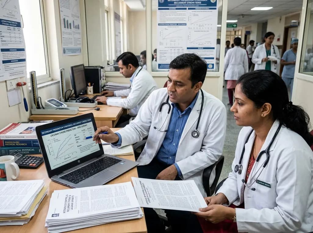
Explore vital recurrent stroke predictors in lacunar stroke patients managed with intensive blood pressure lowering. This analysis examines trial findings from SPS3 to uncover residual vascular risks, metabolic factors, and targeted secondary prevention strategies for high-risk neurological patients.
Today

A comprehensive scoping review synthesises evidence linking prenatal maternal mental health to long-term offspring neurodevelopment, evaluating neuroendocrine, inflammatory, and epigenetic pathways while highlighting clinical screening and future research priorities.
Today
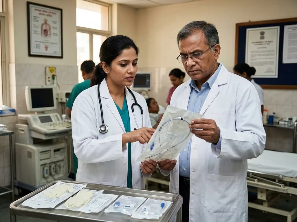
The Directorate General of Health Services directs Indian states to halt the hazardous reuse and reprocessing of single-use medical devices. Discover regulatory implications under the Drugs and Cosmetics Act, critical infection risks, legal liabilities, and operational changes required across clinical specialties.
Today
Mitochondria-targeted nanoparticles self-assembled from EGCG and L-glutamic acid effectively restore mitochondrial homeostasis, suppress synovial inflammation, and attenuate chondrocyte senescence in osteoarthritis, offering a promising disease-modifying nanotherapy.
Today

India is advancing preventive healthcare by integrating genomics, artificial intelligence, and quantum technologies. With the SANGAM programme sequencing one million Indian genomes, clinicians can unlock personalized therapies, detect cardiometabolic risks early, and curb the burden of diabetes and heart disease.
Today
