.webp&w=384&q=75)

Loading, please wait...
Loading, please wait...

Aplasia cutis congenita represents a rare malformation characterized by the focal absence of skin at birth. Although lesions can occur anywhere across the integumentary system, they most commonly involve the scalp vertex. Many affected neonates present with superficial cutaneous ulceration confined to the epidermis and dermis. However, severe defects can penetrate deeply into the galea, cranial bone, and underlying dura mater. Consequently, healthcare providers must evaluate every lesion with extreme vigilance. Because exposed cranial structures risk life-threatening sagittal sinus thrombosis or massive hemorrhage, timely clinical staging remains paramount. Furthermore, clinicians must differentiate isolated defects from broader syndromic conditions like Adams-Oliver syndrome. Therefore, physicians should perform meticulous physical examinations to detect subtle limb or craniofacial abnormalities. While superficial lesions frequently heal uneventfully through epithelialization, deep calvarial defects demand immediate multidisciplinary intervention. Thus, comprehensive neonatal assessment dictates whether conservative management or urgent surgical stabilization offers the safest clinical trajectory. In addition, early parental counseling reduces emotional distress regarding the atypical cosmetic appearance. Consequently, establishing clear expectations regarding wound progression fosters strong trust between families and clinical teams.
A recent 11-year retrospective review from a prominent academic pediatric hospital provides compelling real-world evidence regarding clinical trends. The investigation examined records from 144 patients who received comprehensive treatment between 2012 and 2023. Female patients represented 55% of this pediatric cohort, demonstrating a slight female preponderance. Overall, the average defect size measured 7.05 square centimeters, spanning an expansive range between 0.12 and 96 square centimeters. Crucially, the institutional management strategy correlated directly with initial defect dimensions. Clinicians successfully managed patients presenting with minor lesions averaging 1.87 square centimeters through education and scheduled clinical observation alone, encompassing 42% of cases. Meanwhile, infants presenting with moderate defects averaging 7.14 square centimeters received dedicated local wound care, representing 21% of the cohort. Conversely, surgical reconstruction was reserved for patients exhibiting substantial tissue loss averaging 16.19 square centimeters, which accounted for 26% of treated children. In addition, 12% of patients were lost to extended follow-up. Ultimately, these findings affirm that stratified, conservative approaches effectively resolve the vast majority of smaller congenital defects.
Congenital absence of scalp tissue often accompanies significant systemic comorbidities that require prompt diagnostic recognition. In the reviewed 11-year study, 47% of infants presented with associated morbidities, including 12% harboring internal organ anomalies. Cardiac defects, structural central nervous system anomalies, and limb malformations occurred most frequently across affected children. Consequently, clinicians must perform comprehensive systemic examinations in every newly diagnosed neonate. Midline lesions particularly warrant heightened suspicion because they often overlie cranial vault gaps or abnormal intracranial vasculature. Therefore, physicians should obtain cranial ultrasonography or magnetic resonance imaging to assess skull integrity and delineate superior sagittal sinus anatomy. In addition, echocardiography proves indispensable whenever physicians detect heart murmurs or syndromic facial features. Geneticists also provide critical diagnostic clarity when familial transmission patterns or multiple malformations appear. By identifying occult abnormalities early, clinical teams mitigate systemic risks before initiating procedural interventions. Thus, thorough multidisciplinary screening ensures optimal clinical safety beyond addressing the visible cutaneous lesion. Furthermore, regular developmental assessments help detect subtle cognitive or motor delays during early infancy.
Non-operative therapy represents the primary treatment standard for superficial scalp defects with intact underlying pericranium. In the 11-year pediatric cohort, conservative management successfully addressed approximately 63% of all cases. Specifically, small lesions heal through robust spontaneous granulation and re-epithelialization when providers maintain optimal wound conditions. The therapeutic objective centers on sustaining a clean, moist environment that prevents tissue crusting and desiccation. Therefore, clinicians apply non-adherent dressings coated with bland petrolatum or mild topical antimicrobial ointments like mupirocin. Conversely, healthcare teams avoid silver sulfadiazine in young infants because of systemic toxicity concerns. Furthermore, gentle saline irrigation during dressing changes eliminates debris without disrupting fragile new epithelial cells. Caregivers must also protect the healing scalp from inadvertent mechanical trauma and friction during handling. In addition, pediatric nurses educate parents regarding signs of localized infection, including erythema, induration, and foul discharge. Ultimately, structured conservative wound protocols promote safe secondary intention healing while sparing vulnerable infants unnecessary surgical stress. Consequently, routine outpatient monitoring verifies steady wound contraction until complete re-epithelialization occurs. Moreover, close clinical follow-up ensures rapid identification of any healing arrest.
Although conservative care excels for minor defects, extensive lesions require prompt surgical evaluation to avert fatal complications. Within the 11-year hospital cohort, surgical intervention addressed severe defects averaging over 16 square centimeters. Primarily, reconstructive surgery becomes essential when lesions expose underlying calvarial bone, dura mater, or the sagittal venous sinus. Unprotected dural sinuses carry profound risks of catastrophic hemorrhage, air embolism, and lethal bacterial meningitis. Consequently, surgeons must act decisively to secure durable vascularized soft-tissue coverage over vulnerable structures. Reconstructive modalities depend on defect diameter, pericranial integrity, and local tissue mobility. While skin autografts succeed over intact vascularized periosteum, exposed bone or dura necessitates rotational or transposition scalp flaps. Furthermore, advanced acellular dermal matrices increasingly serve as temporary scaffolds, stimulating granulation tissue over exposed bone before definitive grafting. In addition, surgeons perform delicate flap closure without excessive tension to prevent secondary tissue necrosis. Ultimately, timely surgical reconstruction safeguards high-risk neonates from catastrophic neurological emergencies and provides a foundation for future cranioplasty if needed. Therefore, specialized pediatric surgical teams must tailor each intervention according to anatomical depth and defect geometry.
Aplasia cutis congenita arises from multifactorial disruptions during embryonic skin development, typically within the first trimester. Potential etiologies include intrauterine vascular compromise, amniotic band constriction, localized trauma, and maternal exposure to teratogens like methimazole. Additionally, genetic mutations cause familial forms or syndromic presentations like Adams-Oliver syndrome. While most isolated cases occur sporadically without identifiable causes, genetic evaluation helps rule out underlying syndromic conditions.
Surgical intervention becomes necessary when lesions are extraordinarily large, typically exceeding 15 square centimeters, or involve full-thickness absence of cranial bone and dura mater. These deep defects directly expose vital structures like the superior sagittal sinus, creating severe hazards of fatal hemorrhage, air embolism, or meningitis. In such critical circumstances, reconstructive surgeons deploy local vascularized flaps or dermal matrices to provide rapid and durable coverage.
Yes, underlying calvarial defects frequently undergo spontaneous re-ossification without requiring immediate cranioplasty. Because the infant dura mater and surrounding pericranium retain remarkable osteogenic capacity, gradual bone regeneration typically occurs over several months or years. Consequently, surgeons avoid early bone grafting whenever possible. Clinicians monitor skull healing through serial physical examinations and imaging, reserving formal cranioplasty for persistent, non-healing defects during later childhood.
Disclaimer: This content is for informational and educational purposes only... Refer to the latest local and national guidelines for clinical practice.
References

Read summarized clinical updates, watch expert medical content, and earn CME certifications right from your smartphone.
Open your smartphone camera to scan and install.





A retrospective 11-year review evaluates the management of aplasia cutis congenita in 144 pediatric patients. The study highlights size-based management pathways, ranging from conservative wound care for small lesions to surgical reconstruction for large defects, minimizing severe complications.
Today
A recent Journal of Clinical Oncology study demonstrates that industry payments to oncologists directly sway prescribing behavior. Oncologists who received marketing transfers favored promoted therapies over non-promoted alternatives, highlighting critical ethical concerns for evidence-based cancer practice.
Today
A recent qualitative study examines how college students seek digital sexual health information under persistent uncertainty. Discover the concept of functional trust, the clinical hazards of search abandonment, and evidence-based approaches clinicians can adopt to guide youth toward verified reproductive healthcare.
Today

A randomized controlled trial demonstrates that curated FOAMed videos and podcasts produce superior gains in high-risk ECG interpretation and acute coronary syndrome clinical decision-making compared with print-based materials among interprofessional emergency care teams.
Today

A study reveals that Ginsenoside Rg3 attenuates osteoarthritis progression by modulating the Nrf2-mediated autophagy pathway, reducing chondrocyte apoptosis, and curbing extracellular matrix degradation, offering promising chondroprotective potential.
Today
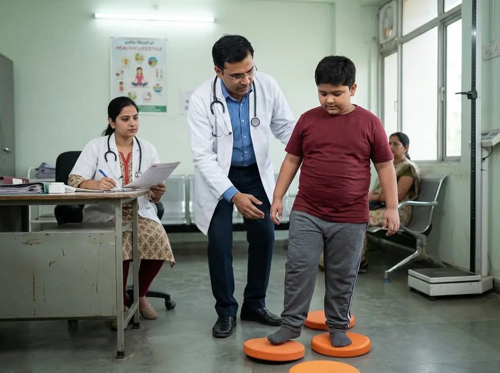
A cross-sectional study demonstrates that lower pediatric motor competence strongly correlates with adverse metabolic syndrome markers, including central adiposity, elevated triglycerides, and low HDL cholesterol, emphasizing early neuromuscular interventions in youth with overweight and obesity.
Today
