.webp&w=384&q=75)

Loading, please wait...
Loading, please wait...

Mammalian oocyte formation is a tightly regulated biological process. It remains essential for female fertility, yet many underlying mechanisms remain poorly understood by the scientific community. To address this gap, researchers recently established an innovative ex vivo culture system. This system faithfully recapitulates in vivo development. Consequently, it enables long-term live imaging of mouse fetal ovaries with unprecedented clarity.
During the study, the team used high-resolution imaging to capture the dynamic behaviors of germ cells. They tracked the development from oogonia into nascent oocytes. Notably, the researchers identified pronounced blebbing activity during the mitosis-to-meiosis transition. Furthermore, meiotic initiation signals strictly regulate this behavior. While the precise role of this blebbing remains unclear, its occurrence underscores its potential developmental relevance in early germ cell maturation.
A prevailing model in reproductive biology suggests that oocyte formation involves organelle transfer. Specifically, the theory posits that organelles move from neighboring germ cells during cyst breakdown to enrich the future oocyte. However, this new study challenges that long-standing assumption. Through advanced photoconversion-based tracking, the scientists observed no detectable transfer of mitochondria or centrosomes. Instead, these organelles remained confined within individual cells.
These findings are significant because they point toward alternative mechanisms for cytoplasmic enrichment. If organelles do not move between cells, the oocyte must accumulate its massive cytoplasmic volume through different pathways. Therefore, this research provides a powerful platform for analyzing germ cell dynamics in real time. It offers a fresh perspective on how the maternal reserve is established before birth.
The study identified intense blebbing during the transition from mitosis to meiosis. While scientists are still determining its exact purpose, the fact that meiotic signals regulate it suggests it plays a key role in early germ cell differentiation.
Contrary to previous models, this research found no evidence of mitochondria or centrosome transfer between cells. The organelles stayed within their original individual cells throughout the observation period.
Disclaimer: This content is for informational and educational purposes only. It does not constitute professional medical advice, diagnosis, or treatment. Always seek the advice of your physician or other qualified healthcare provider with any questions you may have regarding a medical condition. Refer to the latest local and national guidelines for clinical practice.
References
1. Aizawa E et al. Dynamic blebbing and absence of organelle transfer during mouse oocyte formation. EMBO J. 2026 Apr 21. doi: 10.1038/s44318-026-00780-6. PMID: 42014922.
2. Sharma A et al. Oocyte Quality and Female Infertility. Glob J Reprod Med. 2018; 3(2): 555607. DOI: 10.19080/GJORM.2018.03.555607.
3. Lei L, Spradling AC. Mouse oocytes differentiate through organelle enrichment from sister cyst germ cells. Science. 2013; 342(6154): 95-99. DOI: 10.1126/science.1243545.
"
Read summarized clinical updates, watch expert medical content, and earn CME certifications right from your smartphone.
Open your smartphone camera to scan and install.





Research identifies dynamic blebbing and challenges the organelle transfer model in mouse oocyte formation, offering new insights into female fertility....
6 months ago
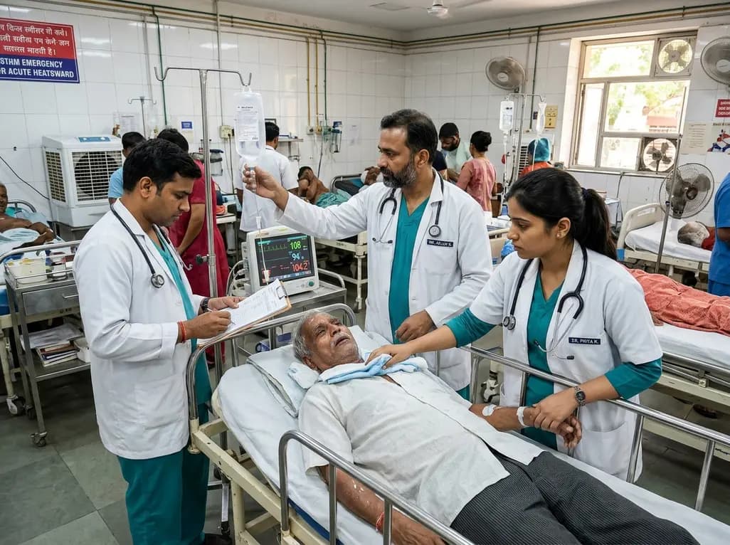
Recent epidemiological data reveals a dramatic surge in summer heat mortality driven by human-induced climate warming. This analysis explores the pathophysiological impacts on cardiac and renal systems, diagnostic dilemmas in death certification, and essential emergency cooling protocols for acute clinical care.
Today
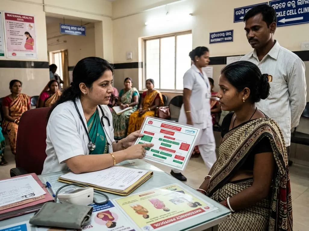
Odisha has established India's first state-level clinical protocol specifically tailored for pregnant women with sickle cell disease. Developed with NHM and UNICEF, the framework provides structured antenatal screening, crisis mitigation, standardized referral pathways, and specialized interdisciplinary care.
Today

India is advancing preventive healthcare by integrating genomics, artificial intelligence, and quantum technologies. With the SANGAM programme sequencing one million Indian genomes, clinicians can unlock personalized therapies, detect cardiometabolic risks early, and curb the burden of diabetes and heart disease.
Today
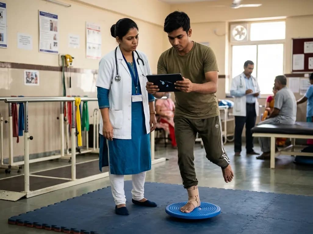
A randomized controlled trial demonstrates that an eight-week dual-task training intervention significantly improves static and dynamic postural control over standard balance exercises in individuals with chronic ankle instability, addressing critical neurocognitive deficits during athletic and daily activities.
Today
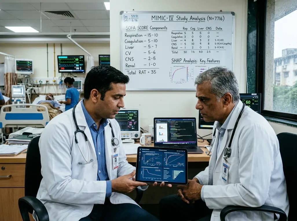
A retrospective cohort study of 7,716 ICU patients from the MIMIC-IV database demonstrates that Gradient Boosting Decision Trees significantly outperform traditional SOFA scores in predicting in-hospital mortality among patients with sepsis and diabetes, with SHAP analysis providing clinical interpretability.
Today
