.webp&w=384&q=75)

Loading, please wait...
Loading, please wait...

Lower-grade gliomas represent a complex group of primary central nervous system neoplasms that frequently affect young and middle-aged adults. Although these World Health Organization grade 2 and grade 3 tumors often present with indolent initial trajectories, they universally carry a devastating biological tendency toward progression. Specifically, malignant transformation in glioma serves as the principal driver of patient morbidity and premature mortality. Clinicians historically struggled to define the precise preoperative determinants and timeline of this catastrophic dedifferentiation. Standard clinical workflows evaluate radiographic changes through intermittent magnetic resonance imaging scans. However, arbitrary surveillance intervals obscure the exact moment a low-grade lesion converts to high-grade malignancy. Consequently, clinicians encounter significant challenges when attempting to accurately stratify risk during initial neurosurgical consultations. Furthermore, conventional predictive models frequently overlook subtle baseline imaging phenotypes. Therefore, identifying robust early markers remains essential for counseling patients and tailoring therapy. By clarifying foundational biological parameters, neuro-oncologists can establish more effective surveillance protocols, ensuring timely interventions before irreversible neurological deterioration occurs.
A major limitation in neuro-oncology literature involves the inaccurate dating of malignant conversion. Historically, investigators recorded the event date as the day a follow-up scan demonstrated overt contrast enhancement or histological progression. However, this approach introduces substantial interval-censoring bias because transformation occurs at an unknown time point between successive scans. Recent methodology rigorously addresses this flaw through interval-censored statistical modeling across complementary analytic frameworks. Notably, researchers demonstrated that conventional event-dating methods overstate the hazard of transformation four-fold. When investigators correct for interval censoring, they reveal a more accurate and nuanced natural history. The study evaluated 155 patients with lower-grade gliomas who demonstrated at most minimal contrast enhancement at presentation. Among non-transforming tumors, surveillance extended over a median of nearly five years. By treating extent of resection as a time-varying covariate, the investigators isolated the true temporal risk profile. Consequently, the corrected models demonstrate that biological progression follows distinct kinetic patterns rather than sudden stochastic shifts. Ultimately, accurate statistical dating provides oncology teams with reliable survival benchmarks and enhances clinical trial design.
The most striking finding centers on diagnostic contrast enhancement. Even when presenting minimally, baseline contrast enhancement represents the single most powerful predictor of malignant conversion. In multivariable models, initial enhancement yielded a Bayesian hazard ratio of 22.1 compared to non-enhancing lesions. Furthermore, this predictive capacity far exceeded the prognostic value of baseline tumor volume. When researchers conditioned the analysis on six-month transformation-free survival, the hazard ratio remained remarkably elevated at 5.65. Most importantly, baseline enhancement predicted post-surgical transformation completely independently of histological grade, IDH mutation status, and surgical resection. Even when neurosurgeons achieved complete macroscopic removal of all enhancing tissue, the baseline signal maintained its prognostic power. This persistent predictive capacity suggests that initial enhancement reflects an intrinsic, aggressive tumor biology rather than mere local vascular disruption. For instance, early microvascular proliferation and blood-brain barrier degradation indicate underlying genomic instability. Accordingly, oncologists cannot assume that removing the enhancing component eliminates the patient's elevated biological risk. Instead, clinicians must treat initial contrast uptake as an indelible biological indicator requiring vigilant long-term surveillance.
While baseline imaging reveals non-modifiable biological risks, surgical resection provides the most powerful modifiable determinant of patient outcomes. The investigation demonstrated that achieving gross-total resection reduces the hazard of malignant transformation by approximately two-thirds. Specifically, complete resection conferred an impressive hazard ratio of 0.32 compared to incomplete debulking or biopsy. Maximizing the extent of resection effectively curtails the residual clonal pool capable of acquiring secondary genetic alterations. Consequently, patients undergoing gross-total resection experience significantly prolonged transformation-free intervals and superior overall survival. However, surgeons often encounter significant technical challenges when operating near eloquent cortex or deep subcortical tracts. In such situations, modern intraoperative adjuncts become indispensable for maximizing resection safely. Neurosurgeons regularly utilize functional cortical mapping, neuronavigation, and intraoperative magnetic resonance imaging to preserve functional integrity while chasing microscopic margins. Furthermore, aggressive cytoreduction reduces local tumor heterogeneity and delays the onset of hypermutation. Therefore, multidisciplinary teams should prioritize maximal safe resection as the foundational therapeutic step. Even in high-risk molecular tumors, aggressive surgical cytoreduction dramatically improves outcomes.
The research team also co-modeled an exhaustive panel of 99 radiomic features alongside traditional radiographic parameters. In recent years, artificial intelligence and texture analyses have generated significant excitement across neuro-oncology. Many clinicians anticipated that complex radiomic signatures would outperform simple visual assessment. Remarkably, no individual radiomic feature survived statistical shrinkage or native interval-censored regression analysis. Simple visual contrast enhancement at diagnosis consistently outperformed the entire computational feature panel. This critical finding provides practical reassurance for clinical teams in resource-limited environments. Sophisticated machine learning algorithms may offer theoretical appeal, but visual assessment on conventional T1-weighted contrast-enhanced magnetic resonance imaging provides immediate, reliable risk assessment. Therefore, clinicians do not need complex radiomic pipelines to identify high-risk individuals. Instead, careful inspection of subtle contrast extravasation guides postoperative surveillance frequency and adjuvant therapy decisions. Because baseline enhancement preserves its prognostic impact following gross-total resection, clinicians must not downgrade vigilance simply because post-resection scans appear clear. On the contrary, these patients warrant intensified radiological surveillance and proactive multidisciplinary review.
Baseline contrast enhancement reflects aggressive intrinsic tumor biology, microvascular proliferation, and genomic instability rather than localized disruption alone. Even when surgeons achieve complete macroscopic resection of enhancing tissue, infiltrative non-enhancing cells retain this high-grade molecular program. Consequently, these residual microscopic populations carry an inherently elevated propensity for subsequent malignant transformation over time.
Standard survival analyses erroneously record malignant transformation at the follow-up scan date, artificially overstating transformation hazard four-fold. In contrast, interval-censored analysis acknowledges that transformation occurs at an unobserved time point between surveillance scans. This rigorous statistical framework eliminates timing biases, providing oncology teams with realistic hazard estimates and accurate natural history timelines.
Gross-total resection serves as the primary modifiable prognostic factor, reducing the hazard of malignant transformation by roughly two-thirds compared to subtotal resection or biopsy. By substantially minimizing the residual tumor cell burden, complete cytoreduction restricts clonal expansion and delays secondary genetic alterations, thereby extending transformation-free survival and improving overall patient outcomes.
Disclaimer: This content is for informational and educational purposes only... Refer to the latest local and national guidelines for clinical practice.
References

Read summarized clinical updates, watch expert medical content, and earn CME certifications right from your smartphone.
Open your smartphone camera to scan and install.





A landmark interval-censored study reveals that contrast enhancement at diagnosis independently predicts malignant transformation in lower-grade glioma, even after gross-total resection. Surgical resection substantially reduces transformation hazard, establishing the primacy of complete tumor removal.
Today
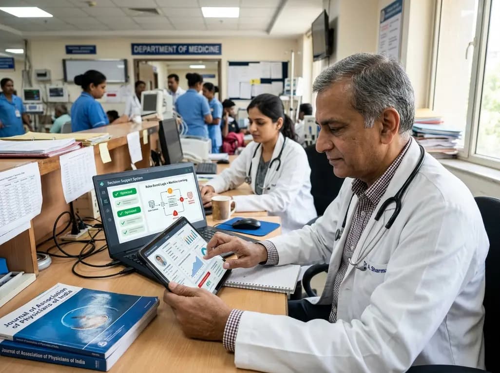
A multicenter study validates a hybrid clinical decision support system combining rule-based logic and machine learning to optimize anticoagulant prescription reviews, reducing alert fatigue and intercepting prescribing errors.
Today

The ClinGen Prenatal Gene Curation Expert Panel evaluated 63 disease relationships across 61 genes, establishing clinical validity for severe fetal phenotypes like hydrops and stillbirth to enhance prenatal genomic interpretation and clinical care.
Today
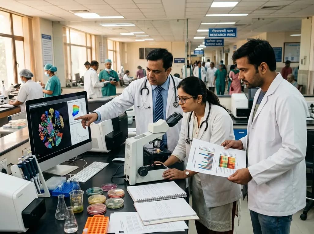
A metataxonomic study reveals distinct gut bacteriome biomarkers in type 2 diabetes, obesity, and cardiovascular complications, identifying specific bacterial shifts that pave the way for precision metabolic medicine.
Today
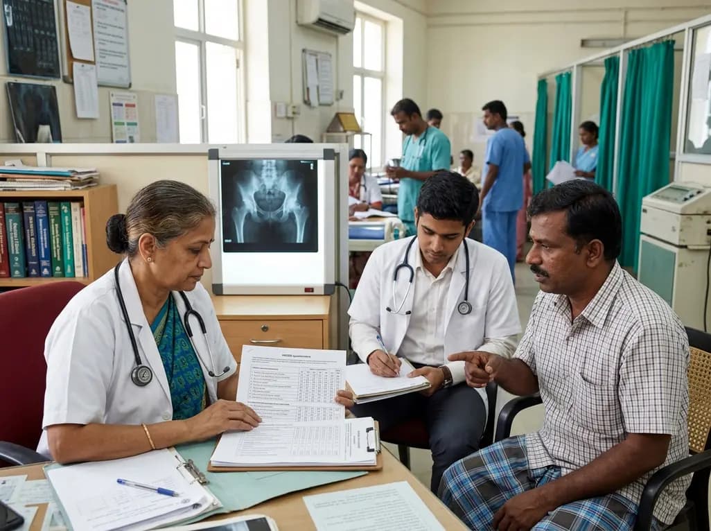
A psychometric validation study confirms that the Turkish Copenhagen Hip and Groin Outcome Score (HAGOS-T) offers excellent reliability, construct validity, and interpretability for patients with hip osteoarthritis, providing clinicians with a robust tool for functional assessment.
Today

Clinical guidelines rely heavily on isolated biomarkers like IGF-1 and HbA1c. However, portal insulin delivery fundamentally gates hepatic growth hormone sensitivity. This physiological continuum unites type 1 and type 2 diabetes, obesity, cirrhosis, and acromegaly, challenging conventional treatment strategies.
Today
