

Loading, please wait...
Loading, please wait...
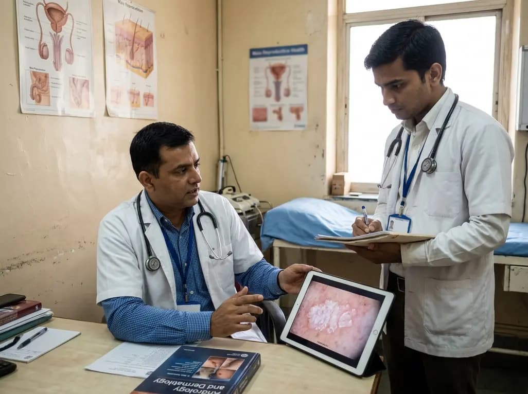
Historically, clinicians managed male genital lesions within isolated surgical siloes, often defaulting to excisional procedures without microscopic clarity. Today, male genital dermatology plays an essential role in bridging the traditional divide between urology and cutaneous medicine. Collaborative diagnostic paradigms now provide vital insights into inflammatory dermatoses, functional complications, and neoplastic risks. Consequently, modern andrological practice relies heavily on dermatological precision to optimize organ preservation, improve surgical timing, and alleviate profound psychosexual morbidity.
For decades, medical literature posited infectious agents or systemic autoimmunity as the drivers of male genital lichen sclerosus. However, robust dermatological investigations refuted roles for human papillomavirus and Borrelia infections. Instead, researchers established chronic occluded urinary exposure as the primary physical instigator of pathology. Micro-incontinence and persistent post-void dribbling trap droplets beneath a tight foreskin, thereby creating chronic cutaneous maceration. Furthermore, this localized toxic chemical irritation incites severe dermal inflammation and progressive microvascular sclerosis. Therefore, clinicians now recognize that uncircumcised anatomy perpetuates this harmful microenvironment. In contrast to female genital disease, circulating autoantibodies rarely influence male penile presentations. Consequently, therapeutic paradigms have moved away from unguided immunosuppression toward mechanical protection and barrier restoration. Physicians clearly appreciate that addressing urinary occlusion remains foundational to halt progressive architectural damage. By redefining the mechanical origins of the disease, male genital dermatology provides a clear rationale for targeted medical regimens and timely operative interventions. Additionally, secondary structural changes like phimosis and frenular tightening exacerbate this fluid entrapment. Therefore, clinicians identify early anatomical compromise before permanent scarring obliterates the urethral meatus.
Accurate identification of benign inflammatory eruptions prevents unwarranted surgical resections. Historically, surgeons classified Zoon's plasma cell balanitis as an autonomous idiopathic disorder. However, recent morphological and clinical evaluations reclassify Zoon's balanitis as a reactive presentation related to male genital lichen sclerosus and chronic prepuce dysfunction. Specifically, persistent heat, moisture, and friction induce dense plasmacytic infiltrates within the superficial dermis. Furthermore, clinicians frequently misdiagnose these shiny, cayenne pepper-like macules as premalignant erythroplasia of Queyrat. Such diagnostic confusion historically prompted aggressive surgical ablation. Today, experienced practitioners employ non-invasive clinical evaluation alongside targeted medical therapies before contemplating surgery. Topical calcineurin inhibitors, including tacrolimus and pimecrolimus, effectively suppress local T-cell responses while restoring epithelial integrity. Moreover, rigorous barrier emollients protect delicate mucosal surfaces from persistent micro-droplet contamination. If medical management fails to relieve symptoms, clinicians execute complete circumcision to eliminate the moist preputial environment. Consequently, patients achieve complete clinical resolution without enduring destructive tissue loss. Collaborative cross-specialty reviews therefore ensure that patients avoid radical treatments for entirely benign reactive dermatoses. Additionally, structured follow-up visits guarantee that clinicians detect subtle recurrences or treatment failures rapidly. This systematic approach ensures optimal penile health and preserves normal sexual function over long clinical courses.
Penile skin presents unique visual challenges due to thin mucosal layers and intense vascularity. Fortunately, the formal introduction of genital dermoscopy has revolutionized chairside evaluation for urologists and andrologists. Clinicians routinely visualize distinctive dermoscopic hallmarks that differentiate inflammatory lesions from early malignancies. For example, male genital lichen sclerosus typically exhibits structureless white areas, keratotic follicular plugging, and subtle linear vessels. In contrast, pigmented penile lesions require rigorous discrimination between benign lentigines, melanosis, and rare malignant melanoma. Dermoscopy readily visualizes pigment patterns, thereby identifying atypical asymmetry and irregular borders with high diagnostic sensitivity. Consequently, andrological clinics have significantly reduced unnecessary, traumatizing penile biopsies. Nevertheless, practitioners maintain decisive biopsy thresholds when lesions present atypical features. Clinicians mandate immediate histopathological sampling for unresolving erythematous plaques, palpable induration, ulceration, or failure to respond to potent topical corticosteroids. Furthermore, expert dermatopathologists interpret these specimen margins with special attention to basal cell architecture. Thus, contemporary dermoscopy establishes an objective triage protocol that prevents both delayed oncological diagnosis and unnecessary anatomical mutilation. Moreover, modern clinics now document high-resolution digital dermoscopic images across successive visits. This structured imaging repository enables specialists to monitor lesion stability with outstanding objective accuracy.
A comprehensive grasp of cutaneous oncogenesis represents one of the most critical contributions of male genital dermatology. Previously, clinicians linked most penile squamous cell carcinomas to oncogenic human papillomavirus genotypes. However, contemporary genomic and histologic research delineates two distinct carcinogenic routes. While classic viral pathways drive undifferentiated penile intraepithelial neoplasia, an independent inflammatory pathway exists. Specifically, male genital lichen sclerosus directly initiates differentiated penile intraepithelial neoplasia. This differentiated neoplastic sequence rapidly advances toward invasive squamous cell carcinoma without viral participation. Consequently, chronic relentless inflammation creates an altered cellular landscape that harbors high rates of TP53 mutations. Urologists must therefore recognize that long-standing genital lichen sclerosus requires perpetual oncological vigilance. Furthermore, pseudoepitheliomatous keratotic and micaceous balanitis represents another elusive entity requiring careful surveillance because it harbors substantial malignant potential. Multidisciplinary tumor boards utilize dermatological expertise to accurately distinguish verrucous carcinomas from extreme reactive hyperplasia. As a result, surgical teams tailor organ-sparing wide local excisions rather than performing radical penile amputations, thereby preserving sexual anatomy and urinary function. In addition, early therapeutic control of mucosal inflammation fundamentally halts neoplastic transformation. Clinicians therefore consider long-term topical surveillance as both a symptom-control strategy and an effective cancer prevention measure.
Effective management of complex penile conditions demands synchronized collaboration between urologists, dermatologists, and reconstructive andrologists. Historically, fragmented care exposed men to conflicting recommendations, ranging from futile topical ointments to excessive distal amputations. Today, integrated penile dermatology clinics combine medical expertise with advanced surgical interventions. For instance, pediatric and adult patients exhibit a triphasic age distribution for lichen sclerosus, necessitating tailored clinical strategies across different life stages. Moreover, previous urological interventions, including catheterisation or hypospadias repair, may provoke koebnerisation and trigger genital dermatoses. Recognizing these triggers prevents clinicians from misinterpreting surgical complications as ordinary postoperative strictures. In addition, chronic penile dermatoses impose an immense psychosexual burden, frequently inducing erectile dysfunction, severe coital pain, and relationship distress. Specialized clinics simultaneously address both cutaneous architecture and psychosexual wellness through targeted medical management, psychosexual counseling, and tissue-sparing surgery. Circumcision remains the definitive curative modality for foreskin-confined disease when topical ultrapotent steroids fail. Ultimately, mutual institutional cross-referrals and joint clinical follow-up secure durable symptom clearance, preserve vital organ function, and dramatically elevate patient quality of life. Furthermore, organized joint clinics create standardized documentation that simplifies long-term surveillance. Consequently, healthcare systems optimize clinical outcomes while minimizing morbidity and costly repeat surgical procedures.
Dermatological investigations confirm that chronic occluded urinary exposure serves as the fundamental physical driver of male genital lichen sclerosus. Micro-incontinence and post-void dribbling allow urine droplets to accumulate beneath an intact foreskin. This persistent occlusion creates cutaneous maceration, compromises the mucosal barrier, and initiates intense localized inflammation. Over time, tissue exposure to urinary toxins triggers progressive sclerosis and scarring. Consequently, relieving occlusion through circumcision or topical management effectively arrests this destructive inflammatory cascade.
Historically considered an autonomous idiopathic disorder, Zoon balanitis is now reclassified as a reactive inflammatory presentation closely linked to male genital lichen sclerosus. Preputial dysfunction, chronic moisture, and trapped urinary secretions induce intense plasmacytic infiltration in the mucosal dermis. Dermatologists recognized that eliminating this moist microenvironment resolves the lesions. Applying topical calcineurin inhibitors or performing curative circumcision halts the plasma cell response completely, eliminating the necessity for aggressive tissue-destructive interventions in affected patients.
Andrologists must order a formal biopsy whenever genital lesions exhibit atypical, progressive, or ulcerated features. Specific clinical indications include persistent erythematous plaques, palpable dermal induration, irregular pigmentary patterns on dermoscopy, or lesions unresponsive to potent topical corticosteroids. Furthermore, suspected differentiated penile intraepithelial neoplasia requires immediate histological confirmation. Prompt biopsy rules out invasive malignancy and guides organ-sparing interventions, ensuring clinicians avoid unnecessary distal amputations while preventing catastrophic diagnostic delays in malignant penile carcinoma.
Disclaimer: This content is for informational and educational purposes only and should not be considered professional medical advice. Always consult with a qualified healthcare provider for specific clinical circumstances. Refer to the latest local and national guidelines for clinical practice.
References

Read summarized clinical updates, watch expert medical content, and earn CME certifications right from your smartphone.
Open your smartphone camera to scan and install.





Male genital dermatology has revolutionized andrology by elucidating the pathogenesis of lichen sclerosus, reclassifying inflammatory balanitis, refining dermoscopic biopsy thresholds, and defining neoplastic pathways to optimize organ preservation and quality of life.
Today

A retrospective cohort study of 7,716 ICU patients from the MIMIC-IV database demonstrates that Gradient Boosting Decision Trees significantly outperform traditional SOFA scores in predicting in-hospital mortality among patients with sepsis and diabetes, with SHAP analysis providing clinical interpretability.
Today

Occult maternal malignancy accounts for 3.82% of persistent unreportable NIPT results with genomic noise. Multiple chromosomal aberrations, notably on chromosome 18, warrant thorough oncological evaluation and whole-body MRI to ensure timely maternal diagnosis while preserving fetal care.
Today

This article reviews the forensic and clinical complexities of acute myocarditis arising on left ventricular hypertrophy in children, highlighting early diagnostic red flags and multidisciplinary postmortem autopsy protocols.
Today

This comparative study evaluates posterior labral tears with and without glenohumeral instability, highlighting distinct demographic patterns, MRI underestimation in stable shoulders, and successful arthroscopic repair outcomes.
Yesterday

A clinical study demonstrates that early intervention with tocilizumab, either as monotherapy or bridged with corticosteroids, achieves significant proptosis reduction and resolves active inflammation in treatment-naive thyroid eye disease.
Yesterday
