

Loading, please wait...
Loading, please wait...
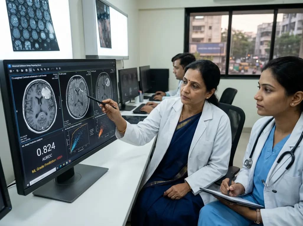
Accurate preoperative meningioma grade prediction represents an urgent unmet need in neurosurgery and neuro-oncology. Intracranial meningiomas constitute the most prevalent primary brain neoplasms in adult clinical practice. Although histopathology reliably defines tumor malignancy after surgical resection, neurosurgeons require noninvasive prognostic insight prior to surgery. Consequently, identifying high-grade tumors before craniotomy allows surgical teams to balance radical resection against the preservation of vital neurological structures. Recently, a major multicenter study published in the Journal of Neurosurgery validated an innovative machine learning model that accurately predicts histological grade using standard diagnostic magnetic resonance imaging.
Historically, clinicians rely on conventional contrast-enhanced magnetic resonance imaging to evaluate intracranial meningiomas. However, qualitative neuroimaging fails to reliably distinguish benign grade 1 tumors from aggressive grade 2 or 3 neoplasms. As a result, operating teams typically discover atypical or anaplastic histology only after completing tissue resection. This diagnostic gap substantially complicates preoperative risk assessment and patient counseling. Furthermore, higher-grade meningiomas demonstrate rapid growth kinetics, extensive bone hyperostosis, and higher recurrence rates. Therefore, surgeons must achieve complete resection, including involved dura, to optimize long-term recurrence-free survival. Conversely, for benign grade 1 meningiomas, surgeons frequently select conservative subtotal resection to protect critical cranial nerves and major venous sinuses. In Indian tertiary care hospitals, neurosurgeons routinely manage complex skull base tumors presenting with substantial mass effect. Consequently, reliable noninvasive grading helps multidisciplinary teams select the safest surgical strategy before craniotomy.
To overcome diagnostic ambiguity, investigators analyzed a large, diverse cohort from the Brain Tumor Segmentation Meningioma Challenge. The study examined multi-sequence imaging data from 909 patients treated across six academic medical centers. Specifically, the authors assigned 699 patients to model development and internal validation, while keeping 210 patients for independent external testing. The image analysis protocol utilized four standard magnetic resonance sequences: T1-weighted, contrast-enhanced T1, T2-weighted, and fluid-attenuated inversion recovery scans. Additionally, expert teams segmented each neoplasm into three distinct physiological compartments, including enhancing tumor, non-enhancing tissue, and peritumoral edema. The automated pipeline subsequently extracted 2,889 radiomic, fractal, and spatial features across these segmented tumor subregions. The investigators systematically evaluated eight machine learning architectures, including deep neural networks, tree ensembles, and linear classifiers. Ultimately, a support vector machine model showed superior performance, providing robust stability and minimal overfitting across diverse institutional scanner protocols.
Through rigorous feature selection, the authors condensed the initial 2,889 parameters into an optimal panel of twenty key features. Importantly, the researchers discovered that conventional radiomic texture metrics alone cannot fully characterize meningioma biology. Traditional radiomics quantifies voxel signal distributions, volumetric shape parameters, and gray-level co-occurrence matrices. However, novel topological data analysis and fractal geometry provided indispensable complementary predictive value. Fractal dimensions measure border irregularity, microvascular tortuosity, and invasive front roughness along the brain-tumor interface. Furthermore, persistent homology captures multi-scale spatial arrangements within both enhancing tumor and surrounding peritumoral edema. Consequently, integrating topological complexity with traditional radiomics significantly improved the algorithm's discriminative precision. This multidimensional mathematical representation reflects underlying histopathological properties, such as high cellular density, sheet-like growth, and elevated mitotic activity. Because these subtle microstructural variations alter radiological patterns, the model successfully identifies aggressive tumor biology long before surgical biopsy.
Many artificial intelligence algorithms demonstrate impressive results during initial internal development but fail when applied to external patient populations. In contrast, the model maintained high diagnostic accuracy on the unseen external validation cohort of 210 patients. Specifically, the model achieved an area under the receiver operating characteristic curve of 0.824, with a ninety-five percent confidence interval of 0.755 to 0.883. Moreover, calibration analyses revealed excellent alignment between predicted probabilities and observed histopathological outcomes. This precise calibration ensures that clinicians can trust the calculated probabilities without fearing excessive false positives or dangerous underestimations. Because the external cohort originated from distinct academic centers using variable magnetic resonance scanners and protocols, these findings confirm generalizability. Thus, the model successfully overcomes the pervasive challenge of inter-scanner domain shift. Additionally, the investigators made the final code and model weights publicly available, enabling worldwide clinical replication and independent verification.
Preoperative grading intelligence directly informs operative decision-making and postoperative surveillance protocols. When algorithmic analysis indicates a high likelihood of grade 2 or grade 3 disease, surgeons can justify aggressive resection strategies. For instance, operating teams may perform radical dural margin excision and hyperostotic bone removal to achieve Simpson Grade 1 resection. Furthermore, surgical teams can anticipate increased vascularity, arrange blood products, and prepare comprehensive cranial nerve monitoring in advance. Conversely, when the algorithm predicts a benign grade 1 tumor, neurosurgeons can confidently prioritize functional preservation. Leaving a thin residual tumor cuff adherent to a critical cavernous carotid artery or optic apparatus preserves life quality without increasing mortality. In Indian neurosurgical centers managing high volumes of complex skull base meningiomas, this individualized risk stratification prevents unnecessary morbidity. In addition, multidisciplinary neuro-oncology boards can schedule early adjuvant radiotherapy for predicted high-grade lesions, avoiding prolonged histopathological delays.
Conventional magnetic resonance imaging relies heavily on visual assessment, which frequently misses subtle microscopic tumor infiltration and cellular heterogeneity. In contrast, machine learning algorithms extract thousands of quantitative radiomic, fractal, and topological features imperceptible to the human eye. This objective computational analysis achieved an external validation AUROC of 0.824. Consequently, machine learning provides standardized, reproducible predictive accuracy that significantly outperforms routine qualitative radiological evaluation alone.
Differentiating benign grade 1 tumors from atypical grade 2 or anaplastic grade 3 meningiomas fundamentally alters surgical strategy. Higher-grade meningiomas exhibit aggressive tissue invasion, rapid growth, and elevated recurrence rates. Therefore, surgeons must pursue radical margin resections, including dural and bone excision, while planning adjuvant radiation therapy promptly. Conversely, for grade 1 lesions, surgeons prioritize cranial nerve and vascular preservation to maintain neurological function.
Yes, because the investigators made the final support vector machine model publicly accessible for clinical and research use. Furthermore, the algorithm requires only routine, multiparametric MRI sequences that hospital radiology suites acquire standardly. Once hospitals automate the multi-region segmentation pipeline, clinicians can integrate the software directly into diagnostic picture archiving systems. This streamlined approach enables rapid preoperative risk stratification without disrupting established clinical workflows.
Disclaimer: This content is for informational and educational purposes only. It is not intended to substitute for professional medical advice, diagnosis, or treatment. Always seek the advice of your physician or other qualified health provider with any questions you may have regarding a medical condition. Refer to the latest local and national guidelines for clinical practice.
References

Read summarized clinical updates, watch expert medical content, and earn CME certifications right from your smartphone.
Open your smartphone camera to scan and install.





A multicenter study in the Journal of Neurosurgery demonstrates an externally validated machine learning model that accurately predicts meningioma grade from preoperative MRI. Combining radiomics and topological features, the support vector machine achieved an AUROC of 0.824 to guide neurosurgical planning.
Today

Gastrointestinal smooth muscle cells rely on nanoscale membrane domains to integrate mechanosensitive currents. Recent insights show that nanodomain architecture, rather than channel properties alone, orchestrates motility, opening new therapeutic avenues for clinical dysmotility and gastrointestinal aging.
Today

A landmark study reveals that lipid-rich malignant ascites alters the T-cell metabolic transcriptome, causing cholesterol efflux and impaired fatty acid oxidation. This suppresses TCR signalling and blocks BiTE immunotherapy, while lipid depletion restores antitumour cytotoxicity in ovarian cancer.
Today
Novel clinical evidence demonstrates the feasibility of combining VA-ECMO with the NyokAssist self-expanding transvalvular pump for active left ventricular unloading in infarct-related cardiogenic shock, achieving successful stabilization and decannulation.
Today
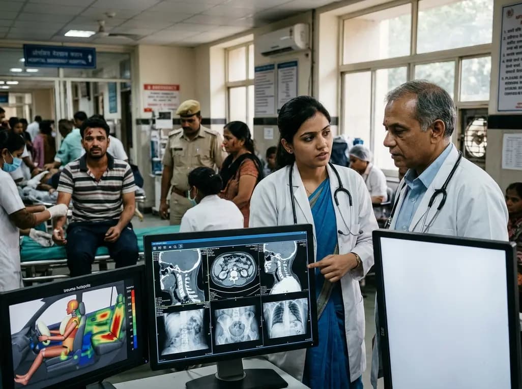
Recent crash analyses demonstrate significant variations in traffic injury severity across vehicle seating positions. Electric vehicle passengers face disproportionately higher trauma risks than drivers, particularly during airbag deployments, demanding adapted emergency assessment and clinical vigilance.
Today
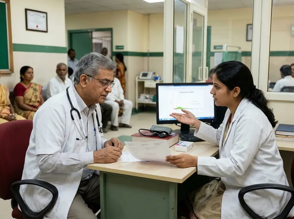
Understand the causal role of Lipoprotein(a) in cardiovascular disease, why international guidelines recommend once-in-a-lifetime molar testing, and the clinical implications of recent phase 3 RNA outcome trials.
Today
