.webp&w=384&q=75)

Loading, please wait...
Loading, please wait...
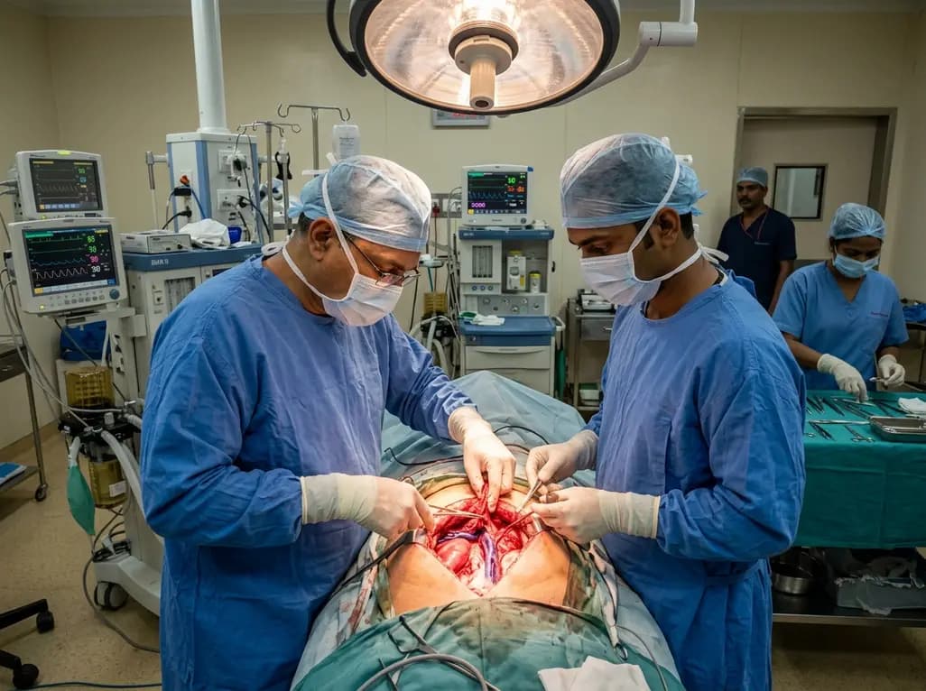
Vascular resection and reconstruction represent essential techniques during radical pancreatectomy for locally advanced pancreatic neoplasms involving the portal-mesenteric venous axis. Surgeons frequently select an autologous left renal vein graft to reconstruct circumferential portal vein defects without creating separate peripheral incisions. Historical evidence suggested that harvesting this vessel carries minimal long-term risk because rich retroperitoneal collateral networks decompress the left kidney. However, clinical observations indicate that postoperative renal impairment occasionally emerges in selected patients following venous harvesting. A landmark study now highlights the critical physiological role of preserving the lumbar vein tributary during left renal vein harvesting. Consequently, understanding venous anatomy and collateral pathways helps surgical oncologists optimize perioperative safety and preserve renal clearance.
The left renal vein occupies a unique retroperitoneal position with extensive tributary architecture. Unlike the short right renal vein, the left renal vein receives venous inflow from three primary collateral structures: the left adrenal vein, the left gonadal vein, and the ascending lumbar vein. When surgeons harvest a left renal vein graft for portomesenteric reconstruction, they ligate the main trunk of the left renal vein near the inferior vena cava. Therefore, renal outflow depends entirely on these three retroperitoneal tributaries to prevent severe venous congestion.
Among these pathways, the lumbar vein provides direct high-capacity communication with the hemiazygos and azygos venous systems. Consequently, the lumbar pathway creates an effective low-resistance outflow channel directly into the systemic circulation. When surgical teams divide or inadvertently sacrifice the lumbar vein during extensive retroperitoneal dissection, collateral decompression becomes severely compromised. Although the left adrenal and gonadal veins offer secondary drainage pathways, their smaller caliber frequently fails to accommodate physiological renal blood flow. As a result, renal parenchymal pressure rises sharply after graft harvest, which ultimately induces microvascular ischemia and impairs glomerular filtration.
A recent clinical investigation evaluated pancreatic resection patients undergoing International Study Group of Pancreatic Surgery type 4 circumferential portal vein reconstruction. The researchers compared renal outcomes between patients receiving a left renal vein graft and individuals reconstructed with alternative vascular conduits. Overall, the general cohort demonstrated comparable glomerular filtration rates across groups. However, granular subgroup analysis uncovered striking functional disparities based strictly on tributary management.
Specifically, the study evaluated patients stratified by lumbar vein preservation versus lumbar vein sacrifice during graft retrieval. Patients in the non-preserved lumbar vein group experienced a significantly higher incidence of postoperative acute kidney injury. Furthermore, these individuals exhibited a persistent median estimated glomerular filtration rate decline exceeding 20 mL/min/1.73 m² at two months postoperatively. In contrast, patients with preserved lumbar tributaries maintained stable renal parameters across postoperative days 3, 7, and 28. Renal scintigraphy confirmed substantial functional depression in the left kidney when the lumbar vein was absent. Interestingly, preserving or dividing the left adrenal vein showed no significant correlation with postoperative renal outcomes.
The pathophysiology of post-harvest renal impairment stems primarily from acute venous hypertension within the renal parenchyma. When surgeons ligate the main left renal vein without preserving adequate collateral outflow, intravascular pressure within the interlobular and arcuate veins surges instantly. This retroperitoneal venous backpressure directly opposes effective renal arterial perfusion pressure. Consequently, the net transcapillary hydrostatic pressure gradient drops, which immediately reduces glomerular ultrafiltration.
Moreover, persistent venous congestion triggers substantial interstitial edema within the rigid renal capsule. This capsular compression collapses peritubular capillaries and produces secondary hypoxic injury to the metabolically active renal tubular epithelium. In addition, cellular swelling and ischemic damage generate localized oxidative stress and inflammatory cytokine release. Although the contralateral right kidney typically compensates over several weeks, the acute bilateral functional burden remains substantial during the critical early recovery phase. For this reason, preserving the high-flow lumbar-azygos axis protects tubular architecture, maintains microvascular perfusion, and prevents severe ischemic acute kidney injury.
Preserving postoperative renal function is crucial for patients undergoing curative-intent resection for pancreatic ductal adenocarcinoma. Standard modern oncological guidelines emphasize early initiation of adjuvant multi-agent chemotherapy regimens, such as modified FOLFIRINOX or gemcitabine-based combinations. However, many cytotoxic agents depend heavily on adequate renal clearance for safe administration and clearance. Thus, prolonged postoperative renal impairment frequently delays or prevents the timely delivery of life-prolonging systemic therapy.
Therefore, surgical teams must rigorously evaluate vascular anatomy before selecting a left renal vein graft for portomesenteric reconstruction. Preoperative contrast-enhanced computed tomography should identify the presence, course, and caliber of the lumbar and gonadal tributaries. If imaging reveals an absent, hypoplastic, or surgically compromised lumbar vein in a patient with borderline baseline renal function, alternative conduits warrant serious consideration. Surgeons can instead choose superficial femoral vein autografts, internal jugular vein grafts, or prosthetic conduits. Consequently, careful preoperative planning aligns surgical vascular reconstruction with downstream medical oncology objectives.
Minimizing collateral disruption requires meticulous dissection technique during graft procurement in the retroperitoneum. Surgeons should expose the anterior surface of the left renal vein between the left gonadal insertion and the inferior vena cava. During this maneuver, the surgical team must exercise extreme caution along the posterior and inferior borders of the vein where lumbar branches typically enter. Gentle vessel loops allow traction without avulsing delicate posterior branches.
Furthermore, surgeons should divide the left renal vein as close to the inferior vena cava as possible while strictly avoiding thermal injury to posterior collateral branches. Hemostatic energy devices must be applied with precise margins to prevent accidental sealing of the lumbar ostium. If retroperitoneal lymphadenectomy necessitates sacrifice of the lumbar system, the operative team should reconsider using the left renal vein. Additionally, maintaining adequate intraoperative hydration and avoiding severe systemic hypotension ensures sufficient driving pressure across the remaining collateral channels. In conclusion, adhering to precise anatomical principles ensures both successful oncological vascular reconstruction and optimal nephrological preservation.
Harvesting the left renal vein disrupts the primary outflow tract of the left kidney, redirecting venous blood through collateral pathways. If surgeons preserve robust collateral tributaries, particularly the lumbar vein, renal function typically remains stable. However, sacrificing these essential collateral channels causes acute renal venous hypertension, leading to significant drops in glomerular filtration rate and increased risks of acute kidney injury.
The lumbar vein connects directly with the extensive, low-resistance hemiazygos and azygos venous systems, enabling efficient systemic drainage under elevated pressure. In contrast, the left adrenal vein possesses a much smaller caliber and limited capacity to accommodate high-volume renal blood flow. Therefore, while adrenal flow provides minimal decompression, the lumbar pathway serves as the primary conduit preventing post-harvest parenchymal congestion.
When the lumbar vein is anatomically absent or compromised during retroperitoneal dissection, surgeons should consider alternative reconstruction grafts. Common autologous options include the superficial femoral vein, internal jugular vein, or great saphenous vein. In addition, cryopreserved allografts or synthetic polytetrafluoroethylene conduits provide reliable vascular reconstruction without risking unilateral renal venous congestion or delaying essential adjuvant chemotherapy.
Disclaimer: This content is for informational and educational purposes only. It is not intended to provide medical advice or to be used as a substitute for clinical judgment. Refer to the latest local and national guidelines for clinical practice.
References

Read summarized clinical updates, watch expert medical content, and earn CME certifications right from your smartphone.
Open your smartphone camera to scan and install.





Preserving the lumbar vein during left renal vein graft harvesting in pancreatic surgery significantly reduces acute kidney injury and long-term eGFR decline, safeguarding renal function for postoperative adjuvant chemotherapy.
last month

A systematic review of 41 preclinical studies reveals that baicalin dampens systemic inflammation, inhibits NF-κB and NLRP3 pathways, and improves organ preservation in sepsis models. However, substantial study heterogeneity and a complete lack of human clinical trials highlight the need for further translational research.
Today
Discover the crucial distinctions between mesonephric and mesonephric-like proliferations of the female genital tract. Learn the key histomorphologic, immunohistochemical, and molecular differences essential for accurate diagnosis and clinical management.
Today

A large UK Biobank study reveals that plasma proteomic signatures capture preclinical organ damage and improve multiorgan risk prediction across early CKM syndrome stages 0 to 2 beyond traditional PREVENT clinical models.
Today
A narrative review investigates whether the articularis genus muscle functions as an independent anatomical entity or blends with the vastus intermedius. We evaluate its morphology, role in retracting the suprapatellar bursa, and direct clinical significance in anterior knee pain and arthroplasty.
Today

A Bayesian multilevel meta-analysis reveals that aerobic training combined with moderate carbohydrate restriction modestly lowers HbA1c in type 2 diabetes. However, sparse data and very low certainty leave incremental benefits over exercise or diet alone unproven, highlighting the need for individualized care.
Today
