.webp&w=384&q=75)

Loading, please wait...
Loading, please wait...
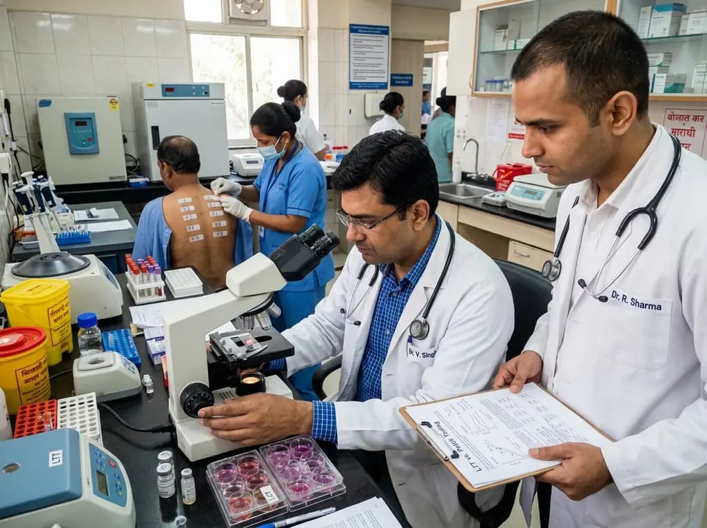
Cutaneous adverse drug reactions remain a pervasive clinical challenge, affecting two to five percent of hospitalized patients worldwide. Specifically, the vast majority of these complications manifest as delayed hypersensitivity drug reactions, presenting predominantly as maculopapular exanthemas. Diagnosing the exact culprit medication creates an intricate puzzle, particularly when patients receive extensive polypharmacy. Clinicians historically struggled to identify inciting compounds without risking severe re-exposure events. Fortunately, modern diagnostic pathways integrate specialized laboratory and cutaneous tests to clarify immune-mediated adverse events. Therefore, evaluating these diagnostic tools enables physicians to optimize therapy, prevent recurrence, and improve overall patient outcomes.
Delayed-type hypersensitivity reactions rely primarily on drug-specific memory T cells that proliferate upon antigen recognition. Therefore, investigators utilize the lymphocyte transformation test to quantify this cellular proliferation in a controlled laboratory environment. The assay measures the uptake of thymidine or metabolic indicators when patient peripheral blood mononuclear cells encounter suspected agents. In recent clinical evaluations, researchers defined a stimulation index of three or greater as a positive threshold. Notably, the test demonstrated an impressive specificity of ninety percent and an equivalent positive predictive value of ninety percent. This high diagnostic accuracy confirms that a positive finding reliably pinpoints the offending agent. Furthermore, the lymphocyte transformation test achieved an overall sensitivity of 64.3 percent across patient cohorts. Consequently, clinicians gain a reliable method to identify immunogenic compounds without administering oral rechallenges. In addition, the assay functions safely because it does not trigger systemic allergic cascades in the patient. Clinicians can confidently isolate culprit drugs while maintaining uninterrupted patient safety.
Patch testing represents an established in vivo technique for detecting delayed cutaneous immune responses. During this procedure, clinicians place non-irritating concentrations of suspected pharmaceutical agents directly onto intact skin using hypoallergenic chambers. Medical teams then evaluate the skin after forty-eight, seventy-two, and ninety-six hours for localized erythema and infiltration. Recent research indicates that drug patch testing achieves an exceptional specificity of one hundred percent and a positive predictive value of one hundred percent. Therefore, any confirmed positive patch result provides conclusive proof regarding the offending agent. However, patch testing demonstrated a modest clinical sensitivity of only 21.4 percent among tested patients. Consequently, a negative patch test cannot rule out drug hypersensitivity. Poor cutaneous penetration and insufficient active drug metabolites often explain these false-negative results. Nonetheless, the procedure remains remarkably safe, accessible, and inexpensive for routine hospital practice. Furthermore, clinicians can perform cutaneous patch testing in ambulatory care settings without specialized laboratories. Accordingly, dermatologists recommend patch testing as a practical initial evaluation for cutaneous drug reactions.
Relying on a single diagnostic modality often leaves clinicians with diagnostic ambiguity during complex clinical investigations. Fortunately, combining the lymphocyte transformation test with cutaneous patch testing significantly expands diagnostic yield. Recent data show that combining both testing modalities raises overall diagnostic sensitivity to 71.4 percent. In clinical studies, patch testing confirmed culprits in 15.8 percent of patients, whereas lymphocyte transformation identified offenders in 44.7 percent of cases. Interestingly, only 7.9 percent of evaluated individuals exhibited simultaneous positive reactivity on both diagnostic tests. Moreover, exactly half of the patient cohort showed negative results across both modalities. These diverse test responses highlight the complementary biological mechanisms governing localized cutaneous absorption and circulating systemic lymphocyte activation. Therefore, integrating both diagnostic tools captures distinct immunological compartments of delayed hypersensitivity. Clinicians effectively reduce diagnostic blind spots by pairing in vitro cellular proliferation with in vivo topical exposure. Consequently, this multi-tiered diagnostic framework empowers physicians to identify culprit medications with enhanced scientific confidence.
Drug classes exhibit substantial variation in their ability to stimulate circulating memory T lymphocytes. Specifically, investigators observed the highest lymphocyte transformation positivity rates for radiocontrast media at sixty percent. Corticosteroids followed closely, demonstrating positive reactivity in 47 percent of tested individuals. Similarly, nonsteroidal anti-inflammatory drugs provoked confirmed cellular proliferation in 43 percent of suspected cases. Although antibiotics constituted almost half of all administered medications, their individual positivity rates remained comparatively lower. In addition to drug classification, specific patient characteristics strongly correlate with test positivity. Notably, a personal history of atopic disease significantly correlates with positive lymphocyte responses. Furthermore, researchers identified the time interval between the initial eruption and diagnostic testing as an essential variable. Specifically, shorter intervals between the cutaneous reaction and laboratory assessment produced significantly higher diagnostic positivity rates. Over time, circulating drug-specific memory T cells naturally decline in peripheral blood. Therefore, clinicians must schedule diagnostic assessments promptly after symptom resolution to capture maximal cellular reactivity.
Inaccurate drug allergy documentation carries severe public health consequences across modern healthcare systems. Patients labeled with unverified allergies frequently receive inferior, more toxic, or markedly more expensive alternative medications. For instance, inaccurate antibiotic allergy labels drive antimicrobial resistance and prolong hospital admissions. Fortunately, high-specificity diagnostic testing provides the objective evidentiary standard required for systematic drug delabeling. Because both patch testing and lymphocyte transformation display outstanding positive predictive values, clinicians can definitively identify true culprits. Consequently, healthcare providers can confidently retain or reintroduce vital non-culprit medications into the patient’s therapeutic record. Moreover, delabeling innocent medications prevents unnecessary lifelong drug avoidance. This objective diagnostic strategy proves particularly advantageous when polypharmacy obscures the primary inciting agent in inpatient wards. In clinical settings across India, where antimicrobials and nonsteroidal agents see extensive use, reliable delabeling protects essential treatment lines. Therefore, structured allergy testing protocols directly improve therapeutic outcomes, enhance antimicrobial stewardship, and protect patient safety.
Clinicians should perform the lymphocyte transformation test between four weeks and six months following complete resolution of the adverse reaction. Testing too early, during acute cutaneous inflammation, may yield false-negative results because circulating reactive T lymphocytes migrate directly into the skin. Conversely, delaying testing beyond several months leads to declining frequencies of circulating drug-specific memory cells. Therefore, scheduling the test within this optimal therapeutic window ensures maximal sensitivity and clinical accuracy.
No, a negative patch test cannot rule out delayed drug hypersensitivity because the test exhibits modest clinical sensitivity. Several factors contribute to false-negative patch results, including poor transepidermal drug penetration, inadequate local vehicle absorption, and the lack of systemic liver metabolism required to produce reactive antigenic metabolites. Consequently, physicians must interpret negative findings cautiously and consider complementary tests, such as lymphocyte transformation assays or supervised drug provocation challenges, before declaring a drug safe.
Non-antibiotic agents like radiocontrast media, corticosteroids, and nonsteroidal anti-inflammatory drugs often trigger distinct immunological mechanisms that elicit robust circulating T-cell responses. Furthermore, certain antibiotics produce unstable reactive intermediate metabolites that research laboratories cannot easily replicate in standard cell cultures. Consequently, non-antibiotic parent compounds can directly stimulate peripheral memory T cells without complex hepatic bioactivation. This biological characteristic produces significantly higher stimulation indices, yielding superior diagnostic detection rates during in vitro lymphocyte transformation assays.
Disclaimer: This content is for informational and educational purposes only... Refer to the latest local and national guidelines for clinical practice.
References
Koumaki D et al. Diagnostic Performance of the Lymphocyte Transformation Test and Patch Testing in Delayed Hypersensitivity Drug Reactions. Dermatitis. 2026 Sep 28. doi: 10.1177/17103568261468627. PMID: 42805914.
Barbaud A. Patch Testing in Drug Eruptions: Practical Aspects and Literature Review of Eruptions and Culprit Drugs. Dermatitis. 2022;33(5):331-344.
Pichler WJ, Tilch J. The lymphocyte transformation test in the diagnosis of drug hypersensitivity. Ann Allergy Asthma Immunol. 2004;93(6):527-535.

Read summarized clinical updates, watch expert medical content, and earn CME certifications right from your smartphone.
Open your smartphone camera to scan and install.





A study evaluates the diagnostic performance of the lymphocyte transformation test (LTT) and patch testing in delayed hypersensitivity drug reactions. LTT demonstrated 90% specificity and 64.3% sensitivity, while patch testing showed 100% specificity. Combining both tests enhances culprit drug identification.
Today

The FEBE study investigates the prevalence of post-traumatic stress disorder symptoms in women receiving high-dose-rate brachytherapy for cervical cancer, highlighting the critical role of procedural sedation and trauma-informed clinical care.
Today

A landmark study reveals that intraoperative goal-directed perfusion targeting oxygen extraction ratio significantly reduces renal replacement therapy and two-year mortality in LVAD explant heart transplantation, offering a vital metabolic target for cardiac surgery teams.
Today

A Level I trauma center study shows a brief bedside mental health intervention boosts 30-day follow-up screening adherence by 84% in nonviolent injury patients. However, outcomes for violence survivors reveal critical care gaps, underscoring the urgent need for specialized community violence intervention programs.
Today
A study introduces a dual-discriminator generative adversarial network (DD-GAN) capable of generating virtual elastography ultrasound from standard B-mode images. The tool matches real elastography performance and improves thyroid nodule risk stratification when combined with TI-RADS.
Today
