.webp&w=384&q=75)

Loading, please wait...
Loading, please wait...

Managing adult patients who experience recurrence after primary glioblastoma treatment presents a major clinical hurdle in modern neuro-oncology. Despite aggressive upfront surgical resection, standardized temozolomide chemotherapy, and localized external beam radiotherapy, almost all high-grade gliomas eventually progress. When treating focal recurrent glioblastoma, clinicians must weigh the risks and therapeutic benefits of repeat interventions. Selecting appropriate salvage techniques requires a nuanced understanding of lesion geography, previously delivered radiation doses, and overall patient performance status.
Patients diagnosed with IDH-wildtype glioblastoma experience rapid tumor recurrence due to the inherently infiltrative biology of these aggressive malignant lesions. Consequently, secondary treatment strategies aim to extend functional independence and provide local cytoreduction without causing debilitating neurological deficits. Open repeat craniotomy offers maximal tumor debulking. However, repeat open surgery often carries elevated morbidity, heightened infection risks, and prolonged hospital stays. Therefore, minimally invasive salvage approaches have gained significant traction among neurosurgeons and radiation oncologists. Specifically, clinicians frequently evaluate laser interstitial thermal therapy and stereotactic radiosurgery as viable local interventions. Both modalities provide targeted cytoreduction while preserving critical adjacent eloquent brain architecture. Nevertheless, comparative evidence clarifying the relative efficacy of these two techniques remains limited. Determining whether one modality confers a distinct survival benefit is essential for tailoring patient-specific salvage regimens.
Understanding the unique biophysical mechanisms of each salvage technique helps clinicians appreciate their respective therapeutic roles. Laser interstitial thermal therapy delivers hyperthermic energy via a stereotactically placed fiberoptic catheter. This localized thermal energy induces immediate coagulative necrosis within the tumor bed. Moreover, magnetic resonance thermal imaging provides real-time monitoring of tissue temperatures, thereby protecting adjacent functional white matter tracts. In contrast, stereotactic radiosurgery administers highly conformal, ablative ionizing radiation doses in single or fractionated sessions. This targeted radiation generates lethal DNA double-strand breaks, which trigger mitotic failure and subsequent tumor cell apoptosis. However, previously irradiated tissues exhibit diminished tolerance to additional ionizing radiation. Consequently, re-irradiation carries a tangible risk of symptomatic radiation necrosis. Conversely, laser thermal ablation operates independently of prior radiation fields. Furthermore, thermal ablation can temporarily disrupt the peritumoral blood-brain barrier, potentially enhancing the central nervous system delivery of concurrent or adjuvant antineoplastic agents.
A recent retrospective investigation evaluated the relative efficacy of these two focal salvage modalities in patients with first-time recurrent IDH-wildtype glioblastoma. The investigation analyzed 57 consecutive adult patients treated between 2010 and 2025, comparing 10 individuals who received laser ablation against 47 individuals who underwent radiosurgical treatment. Notably, univariable survival analysis demonstrated no statistically significant difference in post-recurrence survival between the two cohorts. Patients treated with laser ablation achieved a median survival of 11.5 months, whereas those managed with radiosurgery reached 13.4 months. Because baseline clinical factors can introduce substantial selection bias, researchers performed rigorous multivariate Cox regression modeling and propensity-score matching. The matched cohorts confirmed that survival outcomes remained statistically indistinguishable between the modalities. Therefore, both approaches deliver comparable oncologic efficacy when clinicians apply them to focal high-grade recurrences.
Evaluating procedure-related adverse events is critical when selecting salvage modalities for patients with advanced intracranial malignancies. In the comparative clinical study, investigators observed favorable procedural safety across both intervention groups. The laser interstitial thermal therapy cohort experienced zero direct surgical complications, demonstrating the high precision of modern image-guided stereotactic placement. Conversely, the stereotactic radiosurgery cohort exhibited symptomatic radiation necrosis in 4.3 percent of treated cases. Although radiation necrosis represents an anticipated sequel of high-dose re-irradiation, the low incidence reflects sophisticated conformality in contemporary treatment planning. Additionally, patients who undergo laser ablation typically experience brief hospital admissions and swift functional recovery. Because laser ablation avoids ionizing radiation toxicity, clinicians frequently reserve this technique for patients who have exhausted safe cumulative brain radiation tolerances. Thus, both interventions offer acceptable safety profiles under disciplined multidisciplinary surveillance.
Pre-treatment volumetric characteristics strongly influence procedural selection when managing recurrent high-grade gliomas. In the retrospective analysis, patients chosen for laser interstitial thermal therapy presented with significantly larger baseline contrast-enhancing tumor volumes than those designated for radiosurgery. Specifically, the median recurrence volume was 8.49 cubic centimeters in the thermal ablation group compared to 1.91 cubic centimeters in the radiosurgery cohort. Stereotactic radiosurgery typically demonstrates optimal therapeutic ratios in lesions smaller than two to three centimeters in maximal dimension, as larger target volumes dramatically increase radiation necrosis risks. Conversely, neurosurgeons can effectively deploy laser thermal ablation across moderately larger or irregularly shaped targets by employing multi-trajectory catheter placements. Consequently, lesion geometry, total tumor burden, and distance from eloquent cortical structures dictate appropriate patient allocation. Multidisciplinary neuro-oncology boards should weigh these anatomical factors carefully during therapeutic triage.
The comparable survival metrics observed between laser interstitial thermal therapy and stereotactic radiosurgery underscore the clinical validity of both salvage therapies. Because neither modality demonstrated definitive therapeutic superiority, clinicians can confidently tailor treatment choices to individual anatomical constraints and prior therapy exposures. Moreover, integrating molecular biomarkers and advanced perfusion magnetic resonance imaging will likely refine candidate stratification in subsequent clinical algorithms. Prospective multi-institutional registries and randomized trials are necessary to validate these retrospective findings across larger patient populations. Additionally, combining minimally invasive local cytoreduction with novel immunotherapies or targeted molecular agents represents an exciting therapeutic frontier. Thermal disruption of the blood-brain barrier may facilitate drug delivery, creating powerful combinatorial synergies. Ultimately, neuro-oncologists must continue refining salvage paradigms to extend functional longevity and uphold quality of life.
Current clinical evidence indicates no statistically significant difference in post-recurrence survival between the two modalities. Patients undergoing laser thermal ablation achieve approximately 11.5 months of survival, while those receiving stereotactic radiosurgery achieve approximately 13.4 months. Consequently, both modalities serve as equivalent salvage treatment options for focal recurrence.
Stereotactic radiosurgery works best for small, focal recurrences under two to three centimeters to minimize radiation necrosis. Conversely, laser interstitial thermal therapy accommodates moderately larger or irregularly shaped lesions because neurosurgeons can position multiple trajectories, achieving cytoreduction without delivering additional cumulative ionizing radiation doses to surrounding brain parenchyma.
Laser interstitial thermal therapy carries procedural risks common to stereotactic probes, such as intracranial hemorrhage or localized edema, though complication rates remain low. Stereotactic radiosurgery carries an approximate four percent risk of symptomatic radiation necrosis due to prior tissue irradiation, which clinicians manage using corticosteroids or anti-angiogenic agents.
Disclaimer: This content is for informational and educational purposes only. It is not intended to serve as medical advice, nor does it replace professional clinical judgment, diagnosis, or patient-specific treatment planning. Refer to the latest local and national guidelines for clinical practice.
References
Bindal K et al. Laser interstitial thermal therapy versus stereotactic radiosurgery for first-time treatment of recurrent glioblastoma: a retrospective single-center study. J Neurooncol. 2026 May 19. doi: 10.1007/s11060-026-05632-1. PMID: 42151639.
Montemurro N, et al. Survival outcomes in patients with recurrent glioblastoma treated with Laser Interstitial Thermal Therapy (LITT): A systematic review. Clin Neurol Neurosurg. 2020;195:105942.
Barnett GH, et al. Stereotactic radiosurgery and laser interstitial thermal therapy for recurrent high-grade gliomas: Clinical considerations and patterns of practice. Neurosurgery. 2022;91(5):701-709.

Read summarized clinical updates, watch expert medical content, and earn CME certifications right from your smartphone.
Open your smartphone camera to scan and install.





A comparative study evaluated laser interstitial thermal therapy versus stereotactic radiosurgery for focal recurrent glioblastoma. Both local modalities demonstrated comparable post-recurrence survival, supporting individualized selection based on tumor geometry, lesion volume, and functional anatomy.
Today

Survival analyses often report hazard ratios, but mathematical non-collapsibility creates crucial differences between conditional and marginal estimates. Clinicians must account for unmeasured confounding and model selection when interpreting trial endpoints and real-world cardiovascular outcomes.
Today

A cross-sectional study of 700 college students highlights significant contraceptive knowledge disparities between sexually active and inactive young adults. The findings demonstrate a critical need for proactive, comprehensive sexual health counseling before sexual debut.
Yesterday
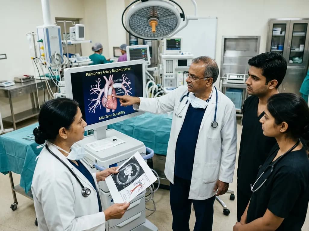
Autologous tissue-engineered vascular graft patches created via in-body tissue architecture show excellent mid-term durability and safety for pulmonary artery reconstruction in congenital heart surgery, particularly for peripheral branch augmentation.
Today

A novel cluster analysis of real-world datasets reveals four distinct CPPD disease phenotypes, validating existing EULAR categories while uncovering previously unrecognized monoarticular and axial presentations to refine patient management.
Today

Recent pharmacokinetic research demonstrates that trace dermal exposure to microgram quantities of trenbolone and metenolone produces detectable urinary metabolites for days to over a week, providing critical objective data for evaluating unintentional contamination claims in sports drug testing.
Today
