.webp&w=384&q=75)

Loading, please wait...
Loading, please wait...
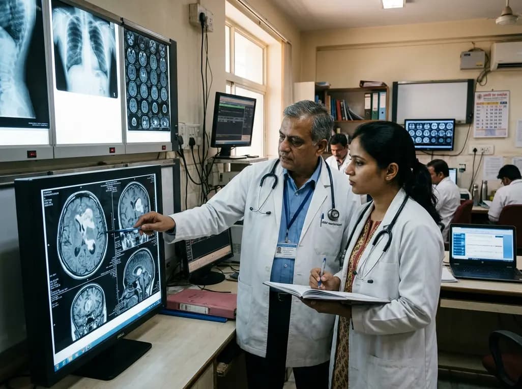
Butterfly gliomas represent one of the most formidable challenges in modern neuro-oncology. These high-grade tumors cross the corpus callosum and invade both cerebral hemispheres symmetrically. Consequently, traditional open surgical resection carries prohibitive risks of severe bilateral neurological injury and profound cognitive decline. Historically, neurosurgeons restricted their intervention to needle biopsy alone. Clinicians subsequently initiated adjuvant chemoradiotherapy without substantial tumor cytoreduction. However, biopsy-only strategies deliver disappointing survival figures, often ranging from only three to six months. Therefore, clinicians urgently seek minimally invasive cytoreductive alternatives. Emerging clinical evidence highlights the utility of LITT for butterfly gliomas to alter the grim prognosis associated with these bilateral lesions. Minimally invasive thermal ablation enables localized tumor cytoreduction while preserving critical callosal pathways and eloquent cortical tracts. As a result, patients can maintain functional independence while accessing standard adjuvant therapies. Recent clinical evaluations demonstrate that aggressive cytoreduction with laser ablation significantly surpasses standard biopsy protocols. Hence, understanding the precise procedural and volumetric dynamics of thermal ablation is essential for neurosurgical oncology teams worldwide.
Laser interstitial thermal therapy offers a stereotactically guided, heat-driven technique for destroying deep-seated malignant tissue. Neurosurgeons place stereotactic laser applicators directly into the core of the lesion through small twist-drill craniostomies. Subsequently, real-time magnetic resonance thermal imaging tracks tissue heating and coagulative necrosis precisely. In addition, integrated cooling systems protect adjacent vital structures from excessive thermal dispersion. This targeted approach enables surgeons to ablate bilateral callosal tumor components without extensive cortical disruption. Furthermore, thermal ablation alters the blood-tumor barrier, which may enhance the penetration of systemic cytotoxic agents. Clinical teams often combine stereotactic biopsy with immediate thermal ablation during a single operative session. Therefore, patients receive definitive tissue diagnosis and meaningful cytoreduction simultaneously. Moreover, the minimally invasive nature of thermal ablation minimizes physical trauma in debilitated cancer patients. As a result, patients experience rapid postoperative mobilization and maintain baseline performance scores. By avoiding wide craniotomies and heavy brain retraction, thermal ablation prevents severe edema and prolonged wound complications. Consequently, this technology reshapes the historical standard of conservative non-resective biopsy for bilateral high-grade gliomas.
Recent longitudinal cohort investigations demonstrate significant survival gains for patients receiving laser ablation over needle biopsy alone. Specifically, clinical research highlights a median overall survival of 14.86 months in ablated patients compared to 4.93 months in biopsy cohorts. Furthermore, median progression-free survival improves significantly, reaching 4.67 months with thermal ablation versus 2.53 months with biopsy alone. These findings confirm that upfront cytoreduction substantially delays tumor recurrence and neurological deterioration. In addition, multivariable analyses indicate that preserving or improving postoperative Karnofsky Performance Scale (KPS) scores directly correlates with extended overall survival. Importantly, patients who maintain their functional independence after ablation tolerate subsequent radiation and temozolomide chemotherapy far better. Accordingly, the hazard ratio for mortality decreases with each incremental point gain in postoperative functional scores. These survival benefits demonstrate that aggressive local cytoreduction fundamentally alters tumor kinetics. Meanwhile, biopsy alone leaves massive active tumor burden, which drives rapid neurological failure and early demise. Thus, offering thermal ablation provides patients with meaningful survival extensions without compromising their day-to-day functional status.
Volumetric analysis provides valuable insights into the mechanical execution and physical limitations of laser ablation. In clinical studies, larger preoperative tumor volumes strongly correlate with lower extent of ablation (EOA) and higher residual tumor burden. Conversely, longer cumulative ablation times allow surgeons to achieve greater volumetric tissue destruction. However, researchers observed an intriguing phenomenon regarding survival metrics. Surprisingly, neither raw extent of ablation nor residual tumor volume directly predicted overall survival in multivariable models. This counterintuitive finding indicates that biological responsiveness extends beyond simple volumetric thresholds. Specifically, coagulative necrosis of hypervascular, metabolically active tumor cores may deactivate critical tumor microenvironments. Moreover, thermal ablation triggers downstream immunogenic cellular responses and disrupts pathological neoangiogenesis. Therefore, neurosurgeons should not view volumetric thresholds as the sole determinant of procedural success. Instead, achieving safe, targeted cytoreduction within deep bilateral tracts provides substantial therapeutic value even when complete ablation remains unachievable. Clinicians must balance maximal thermal delivery against eloquent pathway preservation to ensure optimal clinical outcomes.
Evaluating perioperative safety is essential when comparing minimally invasive thermal therapy to standard needle biopsy. Although total operating room duration is longer for laser ablation (median 4.0 hours versus 2.57 hours), critical safety metrics remain exceptionally favorable. For instance, intensive care unit duration and overall hospital length of stay show no significant difference between ablation and biopsy groups. Furthermore, rates of severe postoperative complications and 30-day hospital readmissions remain equivalent across both cohorts. Consequently, thermal ablation adds profound survival advantages without escalating perioperative morbidity or systemic toxicity. Additionally, patients receiving thermal ablation transition to adjuvant chemoradiation without undue procedural delays. Maintaining this prompt timeline is crucial because delayed radiation therapy correlates with poorer clinical outcomes in glioblastoma. In contrast, extensive open craniotomies frequently induce prolonged wound healing and persistent neurological deficits that postpone adjuvant regimens. Therefore, the favorable recovery kinetics of laser ablation make it an optimal cytoreductive bridge to standard chemoradiotherapy protocols.
The paradigm shift toward minimally invasive ablation requires coordinated evaluation within multidisciplinary neuro-oncology tumor boards. Radiologists, neurosurgeons, medical oncologists, and radiation oncologists must collaborate closely during treatment planning. Early identification of bilateral corpus callosum involvement allows prompt referral to specialized centers equipped with magnetic resonance thermal therapy systems. Furthermore, integrating stereotactic biopsy with immediate ablation eliminates the need for separate diagnostic procedures. Clinicians can subsequently initiate standard-of-care temozolomide and fractionated radiotherapy as soon as the patient recovers. Moreover, future clinical trials are evaluating combined modalities, including laser ablation paired with novel immune checkpoint inhibitors and targeted radiosensitizers. Because thermal damage disrupts the blood-brain barrier, localized delivery of therapeutic agents into peri-ablational zones may enhance long-term disease control. Accordingly, neuro-oncologists should actively consider thermal ablation for newly diagnosed or recurrent butterfly lesions. Replacing passive biopsy strategies with proactive thermal cytoreduction provides tangible hope and measurable clinical benefit for this vulnerable patient cohort.
Butterfly gliomas infiltrate the corpus callosum and spread into both cerebral hemispheres simultaneously. Traditional open surgical resection in these deep, highly eloquent midline structures risks severe neurological impairment, including permanent mutism, hemiparesis, and profound cognitive deficits. Consequently, most centers historically restricted treatment to non-resective biopsy and adjuvant therapies.
Laser interstitial thermal therapy provides direct cytoreduction by causing coagulative necrosis within the tumor core. Recent clinical studies show that thermal ablation nearly triples median overall survival compared to biopsy alone (14.86 versus 4.93 months) and significantly extends progression-free survival without increasing perioperative complications or hospital length of stay.
Preserving and improving the postoperative Karnofsky Performance Scale (KPS) score is the strongest predictor of extended survival following laser ablation. Maintaining functional independence allows patients to complete full adjuvant chemoradiotherapy protocols smoothly, whereas severe baseline deficits and extensive surgical morbidity significantly reduce overall treatment tolerance and longevity.
Disclaimer: This content is for informational and educational purposes only and does not constitute medical advice, diagnosis, or treatment recommendations. Refer to the latest local and national guidelines for clinical practice.
References

Read summarized clinical updates, watch expert medical content, and earn CME certifications right from your smartphone.
Open your smartphone camera to scan and install.





A longitudinal cohort study reveals that laser interstitial thermal therapy (LITT) significantly extends overall and progression-free survival compared to needle biopsy alone in butterfly glioma patients, offering a safe, minimally invasive cytoreductive alternative.
last month

Health authorities in the Democratic Republic of the Congo report unprecedented mortality from the Bundibugyo virus epidemic. Clinicians worldwide must understand the virological profile, atypical hemorrhagic manifestations, early diagnostic protocols, and containment strategies for this evolving health threat.
Today

A nationwide South Korean study shows underreporting of smoking among women dropped from 57.4% to 36.4% between 2008 and 2021. However, over one-third of female smokers still conceal their habit, highlighting the critical role of cotinine verification in clinical risk assessment.
Today
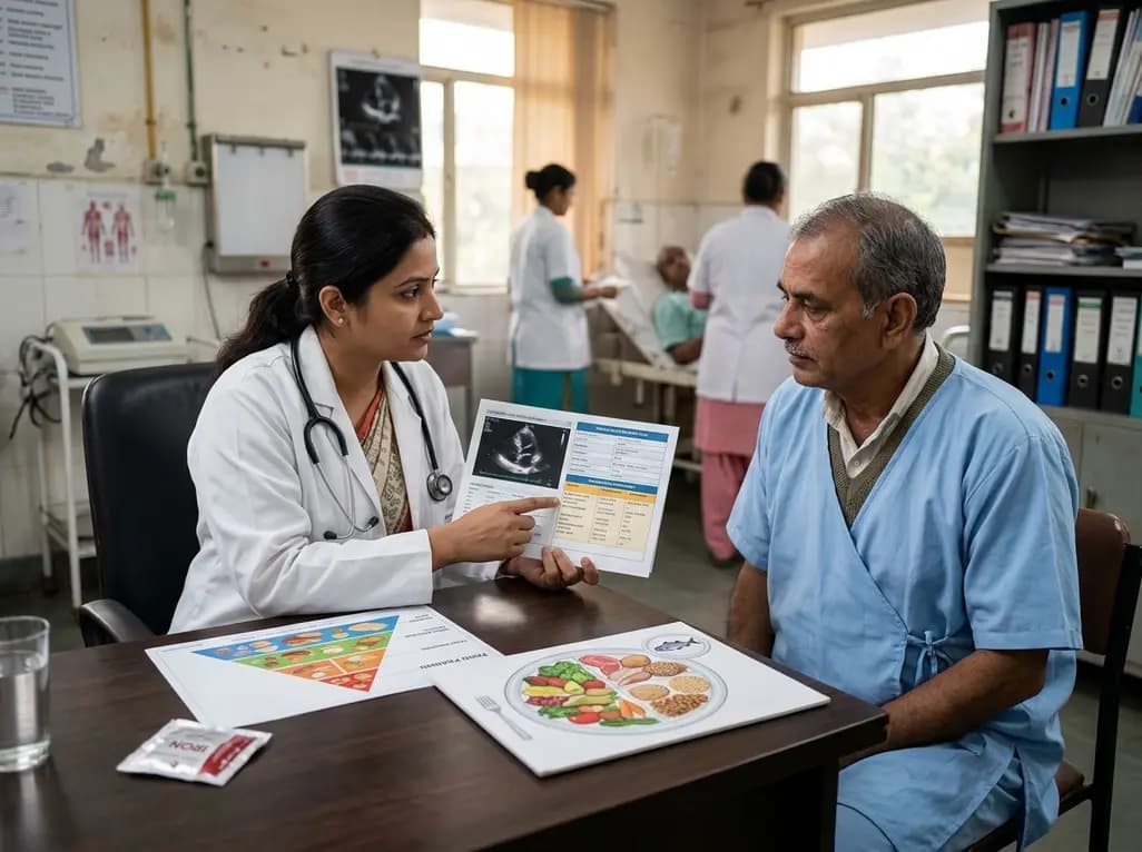
A scientific statement highlights the crucial role of nutrition in managing chronic heart failure. It emphasizes GLIM-based screening, individualized dietary patterns like the Mediterranean diet, appropriate protein and energy intake, avoiding unnecessary fluid restriction, and targeted iron supplementation.
Today

A new study evaluates the rheological stability of decellularized peripheral nerve matrix hydrogels under varied storage conditions. Findings show -80°C storage and freeze-drying preserve gelation kinetics and mechanical stiffness, offering key insights for shipping and translational clinical applications.
Today

A qualitative meta-synthesis highlights the complex interplay between gender dysphoria, minority stress, and eating disorders in transgender and gender-diverse individuals. Clinicians must recognize these distinct drivers to offer supportive, gender-affirming, and interdisciplinary eating disorder care.
Today
