.webp&w=384&q=75)

Loading, please wait...
Loading, please wait...
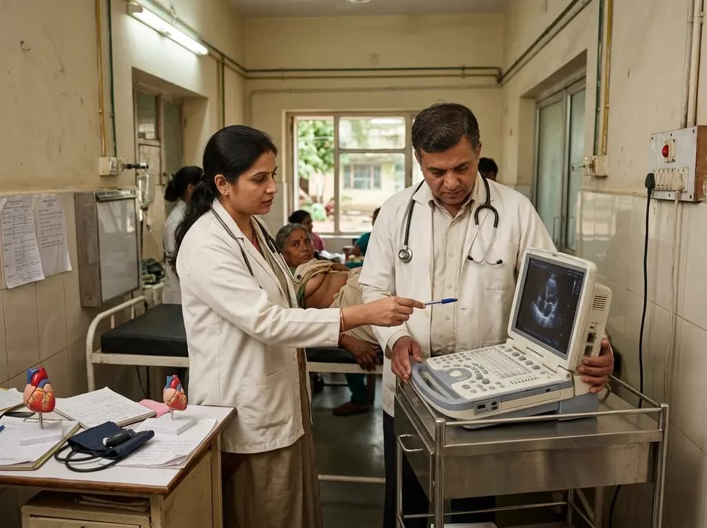
Primary intracardiac neoplasms represent an uncommon yet high-stakes diagnostic category in clinical cardiology. Among these benign neoplasms, a left ventricular myxoma is an exceptionally rare entity that accounts for less than two percent of all cardiac myxoma cases. Most myxomas arise within the left atrium. However, atypical ventricular origins can trigger severe hemodynamic compromise. Clinicians in frontline settings frequently encounter unexplained syncope, which demands a broad and vigilant differential diagnosis. When a mobile ventricular mass occupies the outflow tract, it can cause transient flow limitation and abrupt cerebral hypoperfusion. Therefore, early recognition remains crucial to prevent sudden cardiovascular collapse.
Intracardiac myxomas are histologically benign lesions composed of multipotent mesenchymal cells within an acid mucopolysaccharide-rich stroma. Nevertheless, their anatomical location determines their clinical behavior and risk profile. When a myxoma arises from the left ventricular endocardium, the interventricular septum, or the subvalvular mitral apparatus, systolic flow dynamics push the mass toward the aortic valve. Consequently, this movement causes dynamic left ventricular outflow tract (LVOT) obstruction. The degree of outflow impedance fluctuates with changes in systemic vascular resistance, heart rate, and intravascular volume.
Furthermore, this dynamic obstruction can severely decrease cardiac output during exertion or orthostatic stress, precipitating recurrent syncope. In addition to mechanical flow limitation, mobile intracardiac tumors present a persistent risk of systemic embolization. Fragmented tumor tissue or surface thrombi can dislodge into the cerebral or peripheral circulation, leading to acute ischemic strokes or limb ischemia. Thus, clinicians must recognize that mechanical obstruction and thromboembolism represent dual life-threatening risks in affected patients.
In rural and remote health centers, advanced diagnostic infrastructure is often scarce. Consequently, clinicians must rely on portable technology and fundamental physical assessment skills. Point-of-care ultrasound (POCUS) has emerged as an indispensable bedside modality in primary and secondary healthcare centers. When evaluating a young patient presenting with recurrent syncopal episodes, bedside echocardiography can immediately identify abnormal intracardiac echogenic structures and rule out pericardial effusions.
Moreover, performing a focused cardiac ultrasound enables frontline physicians to detect ventricular wall motion abnormalities, gross valvular dysfunction, and mobile intracardiac masses. While POCUS does not replace a comprehensive echocardiogram, it rapidly shifts the clinical trajectory. Instead of treating recurrent syncope empirically as vasovagal episodes or epilepsy, providers can promptly identify a critical structural lesion. Therefore, integrating POCUS into rural emergency rooms and primary clinics bridges the diagnostic gap and accelerates life-saving specialist consultations.
Following initial detection, patients require structured transfer to secondary or tertiary referral hospitals for comprehensive cardiovascular evaluation. Formal transthoracic echocardiography (TTE) serves as the cornerstone for anatomic and hemodynamic characterization. Specifically, Doppler imaging quantifies peak and mean trans-tumoral gradients across the LVOT, helping clinicians evaluate the severity of obstruction. Color flow mapping also reveals associated mitral or aortic regurgitation caused by mass impingement.
Additionally, transesophageal echocardiography (TEE) provides superior spatial resolution to define the precise tumor pedicle, origin site, and mobility patterns. Contrast-enhanced computed tomography (CT) and cardiac magnetic resonance (CMR) imaging further refine the preoperative assessment. CMR effectively distinguishes myxomas from thrombi, lipomas, and malignant sarcomas using T1- and T2-weighted tissue characterization. Furthermore, multislice CT evaluates coronary anatomy and rules out extracardiac spread or distant embolization. Together, these imaging modalities guide surgical planning and minimize intraoperative complications.
Once clinicians establish the diagnosis of an obstructive ventricular tumor, urgent surgical resection is the definitive treatment of choice. Prompt excision is essential because conservative medical therapy cannot alleviate mechanical obstruction or prevent sudden embolic events. Cardiac surgeons perform complete tumor excision under cardiopulmonary bypass, using median sternotomy with bicaval cannulation to secure optimal exposure.
During surgery, the surgical team prioritizes complete excision of the tumor along with its base to minimize recurrence risks. Surgeons take meticulous precautions to avoid mass fragmentation, which could cause catastrophic perioperative embolic strokes. If the myxoma involves the mitral apparatus, operators perform concomitant valve repair or replacement to preserve left ventricular mechanics. Postoperative recovery is generally favorable, with rapid normalization of intracardiac pressures. However, patients require periodic follow-up echocardiography for several years to monitor for potential tumor recurrence or familial syndromes such as Carney complex.
The successful management of complex cardiovascular disease in underserved regions relies on structured tiered referral systems. Primary care providers, district physicians, cardiologists, and cardiothoracic surgeons must work collaboratively within standardized care pathways. Without efficient regional networking, diagnostic delays can lead to irreversible hemodynamic deterioration or fatal embolic events.
Accordingly, healthcare systems should invest in tele-echocardiography and structured transfer protocols between rural facilities and national centers of excellence. Standardizing emergency referral criteria ensures that patients diagnosed via bedside ultrasound receive timely surgical evaluation. Ultimately, combining point-of-care diagnostics, multidisciplinary collaboration, and robust referral networks significantly reduces mortality from surgically curable cardiac disorders worldwide.
An intracardiac myxoma causes recurrent syncope primarily through dynamic mechanical obstruction of blood flow. When the mobile tumor swings into the left ventricular outflow tract during systole, it abruptly restricts forward stroke volume. This sudden drop in cardiac output reduces cerebral perfusion, leading to transient loss of consciousness, particularly during position changes or physical exertion.
Transthoracic echocardiography serves as the initial diagnostic tool, often preceded by point-of-care ultrasound in emergency settings. Transesophageal echocardiography offers high-resolution visualization of tumor mobility and attachments. Furthermore, cardiac magnetic resonance imaging and computed tomography provide detailed tissue characterization and evaluate hemodynamic compromise before definitive cardiothoracic surgical intervention.
Although cardiac myxomas are histologically benign, prompt surgical resection is mandatory because they pose immediate, life-threatening complications. These hazards include sudden fatal outflow tract obstruction, severe valvular destruction, intractable arrhythmias, and systemic thromboembolism leading to stroke. Surgical excision provides immediate hemodynamic relief and eliminates the risk of future embolization.
Disclaimer: This content is for informational and educational purposes only and should not be considered medical advice. Healthcare professionals should rely on their clinical judgment and verify details independently. Refer to the latest local and national guidelines for clinical practice.
References
1. Murji A et al. Cardiac Myxoma Causing Left Ventricular Outflow Tract Obstruction and Syncope in Rural Tanzania. JACC Case Rep. 2026 Aug 15. doi: undefined. PMID: 42603161.
2. Tyebally S, Chen D, Bhattacharyya S, et al. Cardiac Tumors: JACC CardioOncology State-of-the-Art Review. JACC CardioOncol. 2020;2(2):293-311.
3. Bussani R, Castrichini M, Lionetti V, et al. Cardiac Tumors: Diagnosis and Management. Curr Cardiol Rep. 2020;22(12):169.

Read summarized clinical updates, watch expert medical content, and earn CME certifications right from your smartphone.
Open your smartphone camera to scan and install.





A rare case of left ventricular myxoma causing dynamic LVOT obstruction and syncope highlights the life-saving impact of bedside ultrasound and timely surgical referral in resource-limited settings.
last month
A retrospective clinical analysis reveals that hemodialysis-associated cognitive dysfunction arises from synergistic pathological mechanisms, including severe chronic anemia, worsening renal clearance, abnormal regional brain iron deposition, and an imbalance between neuroinflammatory and neurotrophic factors.
Today

A multiparametric MRI approach combining quantitative T2 shading, apparent diffusion coefficient (ADC) measurements, and morphological signs provides superior diagnostic accuracy over subjective evaluation to differentiate ovarian endometriomas from benign hemorrhagic cysts.
Today

Discover a novel laparoscopic approach for iatrogenic diaphragmatic hernia repair following pedicled omentoplasty. This modified Sugarbaker technique integrates dorsal pedicle lateralization and round ligament reinforcement to secure the diaphragmatic defect while preserving vital omental vascularity.
Today
Evaluating Hill-Sachs lesion size and the distance to dislocation is critical in recurrent shoulder instability. As bone loss concepts shift from binary on-track classifications to continuous risk assessment, clinicians must identify when isolated Bankart repair is insufficient and supplementary procedures are needed.
Today

New research reveals that M1 macrophage-derived exosomes aggravate diabetic nephropathy by transferring WTAP to stabilize S1PR2 mRNA. Silencing WTAP in these vesicles attenuates endothelial injury and renal fibrosis, pointing toward innovative nanomedicine therapies.
Today
