.webp&w=384&q=75)

Loading, please wait...
Loading, please wait...
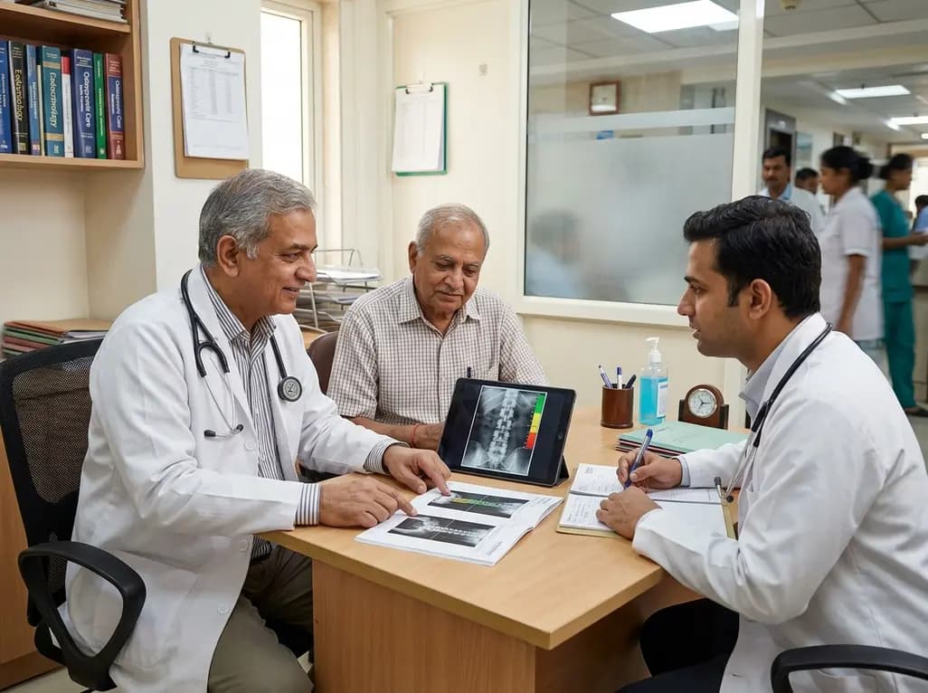
Klinefelter syndrome is the most frequent sex chromosome disorder in men. Although clinicians classically diagnose this genetic condition during adolescence or early adulthood, advancements in prenatal testing now identify many patients much earlier. In contrast, diagnosis in late adulthood remains infrequent but clinically significant. As diagnostic tools evolve, individuals receive structured lifelong surveillance. Consequently, medical focus must shift beyond reproductive issues toward long-term skeletal complications. Optimizing Klinefelter syndrome bone health represents an essential clinical objective across every decade of life.
Bone loss represents a major and well-recognized complication in males with 47,XXY aneuploidy. Primary testicular failure causes severe testosterone deficiency, which directly impairs osteoblast function and accelerates osteoclastic bone resorption. Consequently, reduced circulating androgens diminish cortical thickness and trabecular connectivity. However, low testosterone does not fully explain skeletal deterioration in these patients. In fact, many individuals exhibit significant osteopenia despite exhibiting normal or compensated androgen levels.
Researchers now recognize that global Leydig cell dysfunction produces multi-hormonal deficits. Specifically, decreased insulin-like peptide 3 concentrations impair bone formation and osteoblast differentiation. Furthermore, persistently elevated follicle-stimulating hormone levels stimulate osteoclastogenesis independently of gonadal steroid actions. Genetic dosage effects resulting from the extra X chromosome also perturb androgen receptor gene expression. Additionally, skewed X-chromosome inactivation affects downstream pathways that regulate calcium and phosphate homeostasis. As a result, skeletal microarchitecture deteriorates early, and bone mineral accrual remains compromised throughout young adulthood.
Advancing age substantially alters the clinical trajectory of patients with Klinefelter syndrome. As individuals grow older, biological markers of senescence accelerate, aggravating pre-existing endocrine deficits. In particular, age-related bone loss compounds baseline osteopenia, causing a steep rise in fracture risk. Furthermore, older men frequently develop visceral adiposity and systemic metabolic dysfunction. Insulin resistance, dyslipidemia, and type 2 diabetes mellitus create a persistent pro-inflammatory state that damages bone collagen.
Moreover, chronic cardiovascular disease and progressive sarcopenia often emerge alongside aging in these individuals. Muscle weakness and impaired neuromuscular coordination significantly increase the likelihood of falls, leading to catastrophic fragility fractures. Sedentary lifestyle habits and low physical performance further exacerbate skeletal fragility. In addition, cognitive changes and subtle executive dysfunction may hinder medication adherence and physical safety. Because multi-organ comorbidities accumulate steadily with advancing age, clinicians must monitor older patients vigilantly. Recognizing this multi-system interaction allows physicians to preserve bone integrity before disabling skeletal events occur.
Comprehensive diagnostic evaluation is indispensable for detecting silent skeletal deterioration. Clinicians should perform dual-energy X-ray absorptiometry at baseline for all men diagnosed with the condition. Dual-energy scans should assess the lumbar spine, femoral neck, and total hip to detect localized demineralization. Furthermore, longitudinal dual-energy monitoring helps track progressive bone loss over time. However, conventional areal bone mineral density measurements frequently underestimate true skeletal fragility in this population.
Therefore, physicians should incorporate advanced assessment tools into routine clinical workflows. The trabecular bone score provides critical insight into trabecular microarchitecture without additional radiation exposure. In addition, vertebral fracture assessment and spinal radiographs detect asymptomatic vertebral crush fractures that standard dual-energy scans miss. Laboratory investigations must evaluate serum 25-hydroxyvitamin D, total and free testosterone, gonadotropins, and intact parathyroid hormone levels. Furthermore, clinicians should measure bone turnover markers, such as beta-crosslaps and osteocalcin, to assess remodeling dynamics. Combining these diagnostic modalities ensures accurate fracture risk stratification across all age cohorts.
Prompt therapeutic intervention prevents disabling fragility fractures and preserves overall skeletal stability. Testosterone replacement therapy serves as the primary medical foundation for hypogonadal men. Restoring physiological androgen concentrations stimulates bone modeling and increases lumbar spine bone mineral density. Furthermore, early androgen initiation during adolescence optimizes peak bone mass accrual. However, testosterone replacement alone may not completely normalize cortical geometry or fully eliminate fracture risk in older adults.
Consequently, clinicians must consider targeted bone-active pharmacological agents when severe osteoporosis persists. Antiresorptive agents, such as oral bisphosphonates or intravenous zoledronic acid, effectively suppress excessive osteoclast activity. In addition, subcutaneous denosumab provides a valuable alternative for patients with renal impairment or bisphosphonate intolerance. For high-risk individuals with previous vertebral fractures, anabolic agents like teriparatide rapidly restore trabecular bone architecture. Clinicians must also correct nutritional deficiencies through daily vitamin D and calcium supplementation. Combining hormonal optimization with specific bone antiresorptive therapy provides the most robust skeletal protection in aging patients.
Effective preservation of Klinefelter syndrome bone health requires a continuous, multidisciplinary clinical approach. Endocrinology, urology, geriatrics, and orthopedic specialists must coordinate care to address complex multi-system interactions. Clinicians should prioritize structured lifestyle modifications alongside pharmacological treatments. Specifically, regular weight-bearing exercises and progressive resistance training enhance muscle mass, improve postural balance, and stimulate osteogenesis.
Furthermore, medical teams must educate patients about smoking cessation and moderate alcohol intake to minimize preventable skeletal damage. Regular fall-risk evaluations and home safety modifications protect frail elderly patients against traumatic injuries. In addition, routine monitoring of cardiovascular and metabolic parameters prevents chronic systemic inflammation from worsening bone quality. Clinicians should evaluate mental health, executive function, and treatment adherence during every follow-up consultation. By integrating structured endocrine management with broader geriatric surveillance, healthcare teams ensure prolonged independence and superior quality of life for aging men.
Clinicians should perform a baseline dual-energy X-ray absorptiometry scan at the time of diagnosis, regardless of patient age. Because low bone mass often develops during adolescence, early screening identifies osteopenia before clinical symptoms appear. Clinicians should repeat scans every two to three years in stable patients and more frequently in individuals undergoing active bone therapy or those presenting with uncompensated hypogonadism.
Testosterone replacement therapy significantly improves bone mineral density in hypogonadal men, particularly at the lumbar spine. However, hormonal therapy alone does not always fully normalize skeletal microarchitecture or completely resolve fracture risk. When severe osteoporosis or fragility fractures occur despite adequate androgen replacement, clinicians should promptly combine testosterone with bone-active agents like bisphosphonates, denosumab, or anabolic bone therapies.
Fracture risk in aging men is driven by multiple factors beyond androgen deficiency. Genetic effects from the extra X chromosome, altered androgen receptor sensitivity, and persistent elevations in follicle-stimulating hormone contribute directly to skeletal degradation. Furthermore, age-related sarcopenia, metabolic syndrome, and cognitive changes increase fall frequency, which markedly elevates fracture rates even when circulating testosterone levels appear completely normal.
Disclaimer: This content is for informational and educational purposes only and is not intended to serve as medical advice, diagnosis, or treatment. Healthcare professionals should rely on their clinical judgment and verify all medical information independently. Refer to the latest local and national guidelines for clinical practice.
References

Read summarized clinical updates, watch expert medical content, and earn CME certifications right from your smartphone.
Open your smartphone camera to scan and install.





Klinefelter syndrome requires lifelong surveillance beyond reproductive health. This review highlights how aging exacerbates osteopenia, osteoporosis, and fracture risk, emphasizing early DXA screening, hormonal optimization, and comprehensive multidisciplinary care for older men.
last month

A systematic review of 41 preclinical studies reveals that baicalin dampens systemic inflammation, inhibits NF-κB and NLRP3 pathways, and improves organ preservation in sepsis models. However, substantial study heterogeneity and a complete lack of human clinical trials highlight the need for further translational research.
Today
Discover the crucial distinctions between mesonephric and mesonephric-like proliferations of the female genital tract. Learn the key histomorphologic, immunohistochemical, and molecular differences essential for accurate diagnosis and clinical management.
Today

A large UK Biobank study reveals that plasma proteomic signatures capture preclinical organ damage and improve multiorgan risk prediction across early CKM syndrome stages 0 to 2 beyond traditional PREVENT clinical models.
Today
A narrative review investigates whether the articularis genus muscle functions as an independent anatomical entity or blends with the vastus intermedius. We evaluate its morphology, role in retracting the suprapatellar bursa, and direct clinical significance in anterior knee pain and arthroplasty.
Today

A Bayesian multilevel meta-analysis reveals that aerobic training combined with moderate carbohydrate restriction modestly lowers HbA1c in type 2 diabetes. However, sparse data and very low certainty leave incremental benefits over exercise or diet alone unproven, highlighting the need for individualized care.
Today
