.webp&w=384&q=75)

Loading, please wait...
Loading, please wait...

Acute ischemic stroke remains a major global cause of death and disability. Historically, guidelines restricted intravenous thrombolysis to a narrow therapeutic window of 4.5 hours from symptom onset. Consequently, many patients who presented late lost the opportunity for rapid reperfusion therapy. However, advanced neuroimaging has altered this traditional paradigm. Recent evidence indicates that salvageable ischemic penumbra often persists well beyond the standard timeframe. An updated systematic review and meta-analysis published in Neurology provides robust evidence supporting the IV thrombolysis extended window approach for selected stroke patients, offering renewed hope for delayed presentations.
For decades, acute stroke management relied heavily on the classic "time is brain" paradigm. Clinicians strictly enforced the 4.5-hour cutoff because early clinical trials showed diminishing benefit and rising bleeding risk beyond this boundary. Nevertheless, a substantial proportion of patients present outside this window. Common reasons include wake-up strokes, unwitnessed symptom onset, remote geography, and prehospital transport delays. Consequently, rigid time-based criteria excluded numerous candidates who possessed viable brain tissue.
Fortunately, modern neuroimaging has shifted the focus from time clocks to tissue viability. Contrast CT perfusion and diffusion-weighted magnetic resonance imaging can now distinguish between non-viable core infarct and hypoperfused but salvageable penumbra. Therefore, patients displaying significant mismatch between clinical severity and core infarct size can benefit from thrombolytic recanalization even hours after the initial event. Furthermore, extending treatment windows addresses a critical unmet need in acute emergency stroke care worldwide. Revisiting older restrictions allows clinicians to offer evidence-based reperfusion therapies to individuals who previously received only supportive care.
The newly published systematic review evaluated thirteen randomized controlled trials encompassing 4,867 patients with acute ischemic stroke. Researchers compared intravenous thrombolysis plus best medical therapy against best medical therapy alone beyond 4.5 hours from last known well. The meta-analysis demonstrated clear clinical superiority for the thrombolysis intervention group.
Specifically, patients receiving intravenous thrombolysis achieved a significantly higher rate of excellent functional outcome at 90 days, defined as a modified Rankin Scale score of 0 to 1. The calculated risk ratio was 1.23, corresponding to a number needed to treat of 13. Additionally, secondary efficacy outcomes mirrored these primary findings. Treated patients experienced increased rates of good functional outcome, defined as modified Rankin Scale score 0 to 2, with a risk ratio of 1.15. Moreover, ordinal shift analysis confirmed an overall reduction in disability across all functional levels, yielding a common odds ratio of 1.26. Crucially, trial sequential analysis validated these cumulative efficacy results, proving that the evidence boundary for definitive benefit was crossed successfully.
Safety evaluation remains paramount when administering thrombolytics past traditional cutoffs. The meta-analysis carefully evaluated major safety endpoints, including symptomatic intracranial hemorrhage, total intracranial hemorrhage, and 90-day all-cause mortality.
As anticipated with reperfusion therapies, intravenous thrombolysis was associated with a higher risk of symptomatic intracranial hemorrhage. The study reported a risk ratio of 2.11 for symptomatic intracranial hemorrhage, translating to a number needed to harm of 75. However, this increased bleeding risk did not translate into higher patient mortality. Rates of overall 90-day all-cause mortality were virtually identical between the thrombolysis group and the best medical therapy control group. Similarly, the incidence of any intracranial hemorrhage did not significantly differ between the two cohorts. Ultimately, these safety data indicate that while bleeding risks increase slightly, the substantial gains in functional independence heavily outweigh the numerical risk of symptomatic hemorrhage in appropriately screened stroke populations.
To refine clinical decision-making, the investigators performed detailed prespecified subgroup analyses across several critical variables. Specifically, they examined outcomes based on the choice of thrombolytic agent, extended time window ranges, imaging selection strategies, vascular territory, and concurrent endovascular thrombectomy administration.
Remarkably, no statistically significant subgroup differences were observed. Both alteplase and tenecteplase demonstrated consistent efficacy and safety across extended windows. Furthermore, the clinical benefits persisted whether patients were selected using magnetic resonance imaging or computed tomography perfusion mismatch protocols. Likewise, treatment benefit remained stable regardless of whether patients presented between 4.5 to 9 hours or during wake-up stroke presentations. In addition, the presence or absence of concomitant endovascular thrombectomy did not negate the intrinsic benefit of intravenous thrombolysis. Therefore, these uniform results emphasize that tissue-based selection using advanced imaging serves as a universal predictor of thrombolytic success, irrespective of specific subgroup characteristics or clinical setting.
These comprehensive meta-analytic findings carry significant practical implications for emergency physicians, neurologists, and neurocritical care specialists. Traditionally, late-arriving stroke patients faced limited therapeutic options, leading to severe permanent disability or long-term institutional care. Modern emergency stroke protocols must now adapt to incorporate rapid advanced imaging modalities into routinely available workflows.
Implementing routine perfusion CT or rapid brain MRI in emergency departments enables clinicians to swiftly identify salvageable penumbral tissue beyond the standard 4.5-hour threshold. Consequently, health systems must update acute stroke algorithms, door-to-needle targets, and transfer pathways. Furthermore, establishing clear extended window protocols empowers emergency clinicians to confidently administer thrombolytic therapy to selected patients rather than automatically withholding care due to time delay. Moreover, multidisciplinary care teams, including emergency staff, neuroradiologists, and stroke specialists, must collaborate closely to ensure rapid imaging acquisition and accurate tissue interpretation. Ultimately, integrating extended window thrombolysis into routine clinical pathways can significantly reduce post-stroke disability rates nationwide.
The evolving landscape of stroke reperfusion therapy signals a complete paradigm shift from time-based decision-making to tissue-based personalized care. As robust evidence accumulates, major international and national stroke organizations are updating clinical practice guidelines to endorse extended window thrombolysis for appropriately screened patients.
However, several clinical questions remain active areas of ongoing investigation. Researchers continue to explore optimal dosing regimens for novel thrombolytics, such as tenecteplase, in late-presenting cohorts. Additionally, artificial intelligence algorithms are being developed to automate penumbral mismatch calculations, thereby reducing door-to-treatment times in community hospitals lacking specialized neuroradiology support. Nevertheless, current evidence strongly supports prompt implementation of extended window protocols today. Clinicians no longer need to consider the 4.5-hour clock an absolute barrier to effective pharmacological reperfusion. By prioritizing tissue viability over rigid time windows, medical teams can offer transformative care, restore neurological function, and improve long-term functional recovery for thousands of acute ischemic stroke survivors.
The extended window protocol refers to administering intravenous thrombolytic therapy to selected acute ischemic stroke patients beyond the standard 4.5-hour limit, up to 9 hours or in wake-up strokes. This approach uses advanced imaging like CT perfusion or MRI mismatch to identify salvageable brain tissue before initiating thrombolysis safely.
No, systematic meta-analysis data show that extended window thrombolysis does not increase overall 90-day mortality compared to standard medical therapy. Although the risk of symptomatic intracranial hemorrhage is slightly higher, the overall survival rate remains equivalent while functional independence at 90 days significantly improves in treated patients.
Patient selection relies primarily on neuroimaging demonstrating salvageable brain penumbra rather than exact time elapsed. Clinicians utilize CT perfusion or MRI diffusion-perfusion mismatch to identify hypoperfused tissue that has not yet undergone irreversible infarction. Patients showing significant tissue mismatch without extensive core damage are ideal candidates for extended thrombolysis.
Disclaimer: This content is for informational and educational purposes only and does not constitute medical advice, diagnosis, or treatment. Healthcare professionals should rely on their clinical judgment and refer to the latest local and national guidelines for clinical practice.
References

Read summarized clinical updates, watch expert medical content, and earn CME certifications right from your smartphone.
Open your smartphone camera to scan and install.





A systematic review and meta-analysis published in Neurology demonstrates that IV thrombolysis beyond 4.5 hours from last known well significantly improves 90-day functional recovery in acute ischemic stroke patients selected with advanced imaging, without increasing overall 90-day mortality.
2 months ago
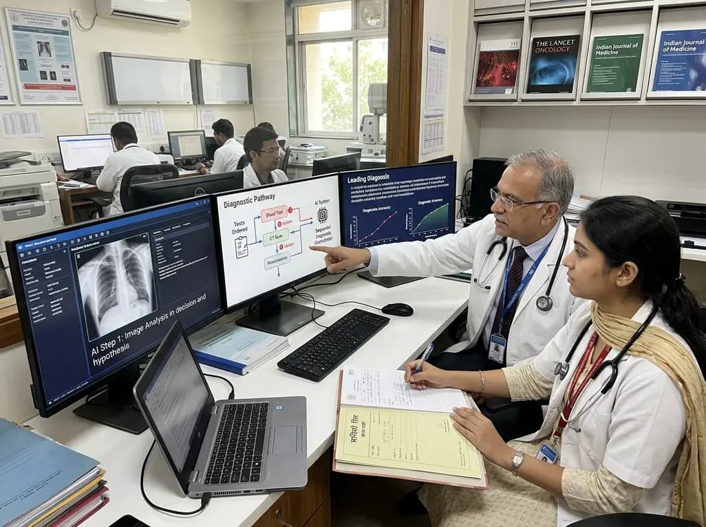
A breakthrough peer-reviewed study demonstrates that a sequential AI diagnosis system can mirror real-world clinical reasoning. By ordering stepwise tests and updating hypotheses dynamically, this multi-agent orchestrator achieved superior diagnostic accuracy while reducing investigative costs.
Today

A prenatal case highlights severe glutaric acidemia type II presenting as bilateral hyperechoic cystic kidneys at 18 weeks. Whole-exome sequencing revealed a novel homozygous ETFDH frameshift variant, demonstrating the power of rapid genetic testing in fetal renal anomalies.
Today

Review how tailored exercise programs benefit frail older patients with heart failure. Explore pathophysiological mechanisms, individualized training prescriptions, and strategies to overcome adherence barriers in clinical practice.
Yesterday

This comparative study evaluates posterior labral tears with and without glenohumeral instability, highlighting distinct demographic patterns, MRI underestimation in stable shoulders, and successful arthroscopic repair outcomes.
Yesterday

A clinical study demonstrates that early intervention with tocilizumab, either as monotherapy or bridged with corticosteroids, achieves significant proptosis reduction and resolves active inflammation in treatment-naive thyroid eye disease.
Yesterday
