

Loading, please wait...
Loading, please wait...
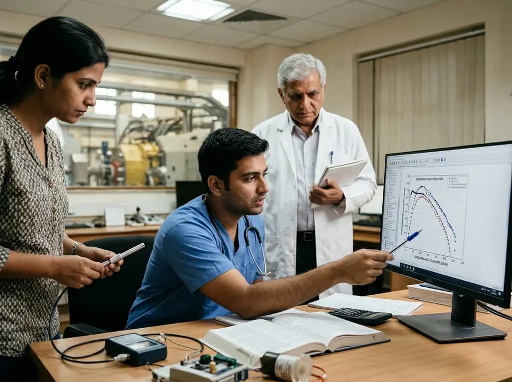
Ultra-high-dose-rate (UHDR) radiotherapy represents a monumental paradigm shift in modern radiation oncology. Preclinical investigations consistently demonstrate that delivering therapeutic radiation at speeds exceeding 40 Gy per second spares normal tissues while preserving tumor destruction. Consequently, this biological phenomenon, recognized as the FLASH effect, attracts substantial clinical interest worldwide. However, synchrotron-based heavy ion and proton accelerators present formidable technological challenges. These facilities deliver beam pulses with intricate microstructural timing and complex spatial profiles. As a result, standard radiation dosimetry struggles to quantify output accurately. Specifically, significant ion chamber recombination occurs within gas-filled measurement cavities under intense instantaneous radiation pulses. When physicists irradiate air cavities at such extreme rates, ion pairs recombine before reaching the collecting electrode. Thus, conventional calibration protocols cannot quantify the true absorbed dose without advanced correction factors. Clinical centers in India, such as proton therapy facilities in Chennai, actively track these developments to evaluate future high-dose-rate applications. Therefore, medical physicists must understand how beam delivery structures influence detector saturation. Addressing these dosimetric hurdles will directly ensure patient safety and beam calibration accuracy before transitioning FLASH modalities into human clinical trials. Moreover, rigorous characterization of particle interactions remains fundamental for establishing reproducible clinical protocols.
Determining the correct dose-rate definition remains essential for modeling ion chamber recombination accurately. Synchrotron beams exhibit substantial temporal variations and sharp spatial gradients. In pencil-beam-scanning delivery, the machine scans small spot beams across target volumes in discrete time increments. Consequently, the dose rate varies by orders of magnitude depending on how investigators define the observation window. Historically, researchers evaluated the field-averaged dose rate across the entire target area. However, this parameter averages exposure across inactive irradiation periods, heavily diluting peak intensities. Similarly, the pencil-beam-scanning dose rate measures spot delivery but often ignores complex spatial overlaps. To resolve these ambiguities, investigators introduced the dose-averaged dose rate. This advanced metric computes a dose-weighted average of instantaneous dose rates across local volume elements. Furthermore, comparative experiments at the Heidelberg Ion-Beam Therapy Center show that this metric consistently predicts higher, more realistic recombination correction factors. Thus, dose-averaged rates align far more closely with experimental observations than alternative parameters. Accordingly, dosimetry teams must adopt this weighted metric during scanned particle therapy beam calibration.
Historically, medical physicists relied on Boag calculations to correct for charge collection inefficiencies in pulsed radiation beams. The classic Boag formulation assumes that radiation pulses deposit energy instantaneously relative to ionic transit times. However, continuous or scanned synchrotron beams defy these simplistic assumptions because beam spills extend over milliseconds. Consequently, researchers formulated quasi-static response models to reconcile dynamic beam delivery with classic physical laws. Under quasi-static conditions, the local dose rate changes slowly compared to the ion transit time across the chamber gap. Therefore, the ionization chamber operates in a state of local instantaneous equilibrium. Nevertheless, when researchers test cylindrical chambers, standard Boag models frequently display noticeable voltage discrepancies. To overcome these limitations, German investigators developed the FLASH Ionization Chamber Model. This specialized numerical transport code models electric field distortions, charge carrier mobilities, and chamber geometries simultaneously. Furthermore, this computational model substantially improves the prediction of voltage dependence compared to standard formulas. Thus, advanced numerical simulations resolve systematic errors caused by simple effective-gap approximations. These computational breakthroughs provide robust foundations for high-precision dosimetry protocols.
Detector geometry significantly influences how ionization chambers behave in extreme radiation environments. At the Heidelberg Ion-Beam Therapy Center, investigators rigorously evaluated three commercial PTW ionization chambers under carbon-ion and proton irradiation. Specifically, the parallel-plate PTW 34045 Advanced Markus chamber demonstrated outstanding performance. Because of its narrow one-millimeter electrode spacing, ions cross the collection volume rapidly. Consequently, the Advanced Markus chamber maintains high charge collection efficiency with minimal ion losses. Similarly, the ultra-small PTW 31023 PinPoint chamber displayed excellent operational stability across both scattered and scanned delivery modes. Its minute sensitive volume effectively limits charge buildup and mitigates volume recombination. In stark contrast, the larger PTW 31009 cylindrical chamber exhibited severe recombination artifacts. The wider cavity volume allowed extensive ion collisions, resulting in substantial deviations between measured charges and model predictions. Furthermore, effective-gap approximations failed to describe the electric field profile in this larger chamber. Therefore, experimental data strongly support the use of thin plane-parallel chambers or miniature cylindrical detectors. By selecting appropriate detector geometries, oncology physicists avoid severe dosimetric underestimations in particle therapy.
The clinical translation of ultra-high-dose-rate particle radiotherapy demands uncompromised beam measurement accuracy. As Indian healthcare institutions invest heavily in advanced cancer technologies, precision dosimetry stands as the cornerstone of treatment quality. Currently, proton facilities in India lead clinical ion beam deliveries in South Asia. In addition, interest in carbon ion therapy continues to grow among national cancer research institutes. If oncology centers adopt ultra-high-dose-rate treatment paradigms, existing quality assurance procedures must evolve rapidly. Conventional reference dosimetry protocols, such as IAEA TRS-398, assume low dose rates and linear recombination corrections. However, applying these standard equations directly to synchrotron-based FLASH fields causes dangerous calibration errors. Therefore, Indian medical physicists must implement dose-averaged dose rate calculations combined with validated transport algorithms. Moreover, radiation oncology teams must establish stringent chamber selection guidelines, favoring detectors like the Advanced Markus chamber. Collaborations between clinical physicists and research laboratories will ensure seamless technological adaptation. Ultimately, implementing rigorous quasi-static models ensures that future Indian cancer patients receive potent particle treatments safely.
Ultra-high-dose-rate beams generate extreme charge densities within detector cavities in microseconds. Consequently, initial and general ion recombination escalate drastically before an external electric field can sweep ions to the electrodes. Standard low-dose-rate assumptions fail because high ion densities heighten the probability of positive and negative ion collisions. Therefore, clinical medical physicists must calculate chamber-specific collection efficiencies to avoid underestimating true absorbed radiation doses during fast FLASH delivery.
Conventional dose-rate metrics, such as field-averaged dose rate, dilute intense local delivery across total exposure time and field dimensions. In contrast, the dose-averaged dose rate weights instantaneous local dose rates against local deposited dose. Thus, it captures steep micro-temporal and spatial delivery peaks occurring during pencil beam scanning. Consequently, this weighted metric closely mirrors physical ion generation inside the chamber cavity, yielding accurate recombination correction factors under quasi-static conditions.
Plane-parallel chambers with narrow electrode separation, notably the PTW 34045 Advanced Markus, demonstrate optimal performance and minimal ion loss. Additionally, ultra-small cylindrical chambers like the PTW 31023 PinPoint perform reliably because their small collection paths mitigate volume recombination. In contrast, larger cylindrical chambers experience severe charge recombination and deviate significantly from standard analytic models. Therefore, physicists prefer small-volume or plane-parallel geometries when calibrating ultra-high-dose-rate particle beams in modern clinics.
Disclaimer: This content is for informational and educational purposes only... Refer to the latest local and national guidelines for clinical practice.
References
Charuchinda W et al. Dose-rate metrics for ionization-chamber recombination in synchrotron-based ultra-high-dose-rate proton and carbon-ion beams: application and validity of quasi-static response models. Z Med Phys. 2026 Nov 07. doi: undefined. PMID: 42998949.
Di Martino F, et al. High dose per pulse electron beam dosimetry - A model to correct for the ion recombination in the Advanced Markus ionization chamber. Med Phys. 2017;44(3):1157-1167.
Romano F, et al. Ultra-high dose rate dosimetry: Challenges and opportunities for FLASH radiation therapy. Med Phys. 2022;49(7):4912-4932.

Read summarized clinical updates, watch expert medical content, and earn CME certifications right from your smartphone.
Open your smartphone camera to scan and install.





This study evaluates dose-rate metrics for ion chamber recombination in synchrotron UHDR proton and carbon-ion beams, highlighting the dose-averaged dose rate (DADR) and quasi-static numerical models for accurate FLASH radiotherapy dosimetry.
Today

A case report details how reusing single-use insulin cartridges caused device failure and diabetic ketoacidosis in a type 1 diabetes patient using an advanced hybrid closed-loop system. The report highlights technological blindspots, mechanical degradation, and safety protocols during supply shortages.
Today

Maternal cardiovascular risk factors such as hypertension, obesity, and diabetes significantly elevate offspring cardiovascular disease risk through placental dysfunction and fetal programming. Enhancing preconception screening and antenatal care offers clinicians a powerful strategy to protect long-term heart health.
Today

New phase I data evaluate the impact of food and beverages on enlicitide, the first oral PCSK9 inhibitor. While food reduces peak absorption, model simulations demonstrate robust LDL-C lowering of approximately 60%, confirming meaningful pharmacokinetic forgiveness and flexible real-world administration.
Yesterday

A retrospective cohort study reveals that lumbar spine bone mineral density does not distinguish extremity long bone fracture status in adults with severe motor and intellectual disabilities. Because age drove fracture risk rather than spinal density, clinicians must adopt comprehensive assessment tools.
Today

A landmark UK Biobank study by Karolinska Institutet researchers reveals that optimal rest mitigates neurodegeneration in cardiometabolic illness. Patients maintaining high sleep quality demonstrated superior cognitive scores and larger brain volumes, highlighting sleep optimization as an actionable clinical strategy.
Yesterday
