

Loading, please wait...
Loading, please wait...

An involuntary inspiratory sigh represents a unique respiratory manifestation that frequently challenges movement disorder specialists during routine clinical evaluations. In patients with multiple system atrophy, this distinctive respiratory pattern functions as an established supportive diagnostic feature. However, clinicians often overlook its occurrence during wakefulness because daytime respiratory assessments remain inconsistent in standard clinical practice. Multiple system atrophy is an aggressive, progressive neurodegenerative illness characterized by autonomic failure, parkinsonism, and cerebellar ataxia. Respiratory disturbances, including daytime sighs, stridor, and sleep-disordered breathing, contribute significantly to patient morbidity and sudden nocturnal mortality. Although sleep-related sighs have received academic interest, daytime involuntary sighing provides immediate bedside clues to underlying neurodegeneration. Consequently, recognizing this specific sign allows neurologists to differentiate multiple system atrophy from classic Parkinson's disease early in the illness course. When healthcare practitioners evaluate unexplained atypical parkinsonian symptoms, active observation of daytime breathing irregularities yields indispensable diagnostic insight. Therefore, understanding the presentation, frequency, and clinical implications of this breathing abnormality is vital for contemporary neurological care.
Recent clinical investigations demonstrate that involuntary inspiratory sigh correlates directly with greater overall disease severity in multiple system atrophy. In a systematic cohort study of ninety patients, researchers identified this respiratory sign in nearly one-quarter of all confirmed cases. Patients exhibiting daytime sighs consistently presented with significantly higher scores on Part I of the Unified Multiple System Atrophy Rating Scale. This rating scale specifically reflects impairment in historical activities of daily living, indicating extensive functional disability among affected individuals. Furthermore, these patients demonstrated higher scores on the rapid eye movement sleep behavior disorder screening questionnaire. This finding emphasizes that widespread neurodegeneration spans both daytime respiratory motor control and nighttime sleep architecture. As the neurodegenerative pathology expands across subcortical networks, affected individuals experience a compounded functional decline that diminishes quality of life. Moreover, the simultaneous occurrence of severe sleep dysfunction and daytime respiratory abnormalities signifies accelerated disease progression. Consequently, clinicians should consider daytime sighing not merely as an isolated respiratory oddity, but rather as a clinical biomarker reflecting pronounced cumulative motor and non-motor disability.
Autonomic failure constitutes a cornerstone of multiple system atrophy pathology, and respiratory symptoms closely mirror this underlying autonomic breakdown. In exploratory cohort analyses, patients with involuntary inspiratory sigh demonstrated a significantly higher frequency of severe orthostatic hypotension. Orthostatic hypotension poses substantial hazards, including recurrent syncope, debilitating falls, traumatic injuries, and rapid loss of ambulatory independence. The autonomic nervous system intricately regulates cardiovascular reflexes alongside involuntary respiratory drive through dedicated pontomedullary networks. When neurodegeneration ravages central autonomic control centers, both vasomotor tone and respiratory rhythm generators undergo profound disruption. Therefore, the emergence of daytime sighing often alerts clinicians to advanced dysautonomia requiring urgent therapeutic intervention. In daily clinical management, healthcare providers must aggressively screen for postural drops in blood pressure whenever they detect atypical respiratory signs. Addressing orthostatic intolerance through hydration, fludrocortisone, midodrine, or compression garments mitigates injury risk in these highly vulnerable individuals. Ultimately, recognizing this clinical link ensures that physicians deliver timely cardiovascular and supportive autonomic care before catastrophic syncopal episodes occur.
Investigating the neural substrates of involuntary inspiratory sigh reveals essential insights into brain perfusion changes in multiple system atrophy. Single-photon emission computed tomography studies examining cerebral blood flow demonstrate prominent perfusion abnormalities in sigh-positive patients. Specifically, neuroimaging reveals significantly reduced regional cerebral blood flow localized to the bilateral supplementary motor area. The supplementary motor area coordinates complex motor sequencing, postural adjustments, and intentional movement initiation across diverse physiological systems. Additionally, this cortical region communicates extensively with subcortical and brainstem nuclei that regulate automatic motor patterns and respiratory pacing. Reduced perfusion in the supplementary motor area suggests that cortical motor dysfunction contributes directly to aberrant daytime respiratory control. Thus, the clinical sign reflects not only brainstem degeneration but also broader cortical-subcortical network failure. Understanding these functional neuroimaging correlates deepens our grasp of multiple system atrophy pathogenesis. Furthermore, identifying these specific perfusion deficits confirms that respiratory signs arise from distinct neuroanatomical disruptions rather than psychogenic or non-specific behavioral factors.
Detecting respiratory signs during outpatient appointments presents practical logistical challenges for busy healthcare practitioners. In clinical evaluations, researchers compared structured patient interviews against direct bedside examinations to determine the most effective detection strategy. Interestingly, the investigation revealed only moderate agreement between structured caregiver interviews and direct visual examinations conducted by clinicians. Involuntary sighs occur intermittently throughout the day, so short clinical consultations frequently miss these transient events entirely. In contrast, comprehensive structured interviews with patients and primary caregivers captured involuntary inspiratory sigh significantly more reliably. Family members observe subtle breathing changes across prolonged periods at home, making their detailed history an indispensable diagnostic instrument. Therefore, clinicians should not rely exclusively on brief physical observations in the examination room. Instead, physicians must incorporate targeted screening questions about sudden daytime deep gasps or noisy inhalations into routine clinical intake protocols. Actively querying family members ensures timely detection, enabling accurate stratification of disease severity and prompt supportive management.
In India, diagnosing atypical parkinsonian syndromes poses notable clinical challenges due to heavy outpatient volumes and limited access to specialized neuroimaging. Patients with multiple system atrophy are frequently misdiagnosed with idiopathic Parkinson's disease during early symptomatic phases, leading to delayed multidisciplinary management. Incorporating a routine inquiry regarding involuntary inspiratory sigh provides an inexpensive, highly practical clinical tool for Indian neurologists and internists. Because structured interviews reliably reveal this sign, doctors do not require expensive diagnostic machinery to uncover vital prognostic clues. Furthermore, identifying this manifestation should immediately prompt aggressive cardiovascular screening for neurogenic orthostatic hypotension and nocturnal sleep studies to detect laryngeal stridor. Timely identification enables clinicians to initiate early palliative support, fall prevention strategies, and tailored caregiver education. Primary care physicians across district hospitals and tertiary neurology centers can readily adopt this screening approach to enhance diagnostic precision. By paying meticulous attention to respiratory nuances, healthcare providers can dramatically improve symptom management and patient safety across resource-diverse clinical settings.
An involuntary inspiratory sigh is an abnormal respiratory manifestation characterized by sudden, unintentional deep breaths or audible daytime gasps. In multiple system atrophy, it reflects underlying degeneration within central autonomic and motor control circuits. Clinicians consider it a supportive non-motor diagnostic feature that signals higher disease burden and extensive neurological impairment.
Direct clinical examinations frequently miss involuntary sighs because these respiratory events occur intermittently throughout the day during wakefulness. Brief clinical consultations cannot capture sporadic breathing irregularities reliably. Structured interviews with patients and primary caregivers prove significantly more effective, as family members observe the patient over extended periods in home environments.
Upon identifying involuntary sighs, clinicians must promptly conduct formal postural blood pressure evaluations to screen for severe orthostatic hypotension. Additionally, practitioners should assess nighttime respiratory patterns for laryngeal stridor or sleep apnea. Comprehensive multidisciplinary management, aggressive fall prevention counseling, and caregiver education should begin immediately to minimize complication risks.
Disclaimer: This content is for informational and educational purposes only. It is not intended to be a substitute for professional medical advice, diagnosis, or treatment. Always seek the advice of your physician or other qualified health provider with any questions you may have regarding a medical condition. Refer to the latest local and national guidelines for clinical practice.
References

Read summarized clinical updates, watch expert medical content, and earn CME certifications right from your smartphone.
Open your smartphone camera to scan and install.





New clinical research demonstrates that involuntary inspiratory sigh in multiple system atrophy marks higher disease severity, severe orthostatic hypotension, and reduced supplementary motor area perfusion.
Today

A 60-year-old woman presented with subacute cerebellar ataxia, dysautonomia, and hyperreflexia. Despite negative commercial antibody panels, tissue-based assays and phenotypic evolution to blepharospasm guided diagnostic resolution through specialized neuroimmunology testing.
Today

A prospective study reveals significant first-trimester trace-element alterations in pregnant women with epilepsy. Lower serum selenium independently predicted subsequent seizure recurrence despite prior remission, highlighting the vital clinical role of micronutrient profiling during antenatal care.
Today
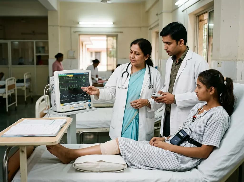
A 15-year-old patient presented with knee arthralgia and acute rheumatic fever, exhibiting complex arrhythmias including AV blocks and non-sustained ventricular tachycardia despite lacking severe valvulitis. This highlights the importance of extended Holter monitoring in pediatric rheumatic carditis.
Today
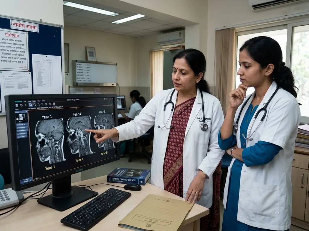
An eight-year longitudinal imaging follow-up reveals severe temporomandibular joint remodeling in nodulosis-arthropathy-osteolysis (NAO) syndrome. Despite extensive osseous destruction, the patient maintained functional jaw opening without TMJ interventions, highlighting the role of conservative management.
Today

Discover essential clinical insights into AGLP1-type amyloidosis, a rare localized complication of glucagon-like peptide-1 analog injections. Learn how histopathology and mass spectrometry differentiate benign iatrogenic amyloidomas from systemic disease, preventing misdiagnosis and unwarranted therapies.
Today
