

Loading, please wait...
Loading, please wait...

Subacute and chronic subdural hematomas represent an increasingly common neurosurgical dilemma among aging populations worldwide. Conventional treatment typically relies on formal operating room interventions, such as burr hole drainage or twist-drill craniostomy. However, geriatric patients frequently present with pronounced frailty, severe cardiovascular comorbidities, and concurrent anticoagulant therapy. These complex medical conditions significantly heighten perioperative mortality and anesthetic complications. Consequently, surgical teams require less invasive bedside approaches that maintain patient safety while achieving rapid cerebral decompression. Emerging clinical evidence highlights bedside IO needle decompression as an effective, minimally invasive therapeutic solution. By utilizing standard intraosseous vascular access devices, clinicians can evacuate subdural collections without requiring general endotracheal anesthesia. Furthermore, this innovative bedside intervention provides immediate intracranial pressure relief directly in the emergency department or intensive care unit. As neurotrauma cases continue to rise globally, understanding this procedural technique broadens clinical options for acute care teams.
Chronic subdural hematoma predominantly affects older adults following mild head trauma. Over several weeks, bridging cortical veins tear and initiate slow intracranial hemorrhage. An persistent inflammatory cascade subsequently develops, promoting fragile neocapillary growth and recurrent microbleeding within the subdural space. Consequently, the hematoma gradually enlarges and exerts considerable mass effect on adjacent brain parenchyma. Patients frequently present with progressive gait ataxia, insidious cognitive decline, persistent headache, or localized motor deficits. In emergency settings, clinicians often misdiagnose these non-specific manifestations as ischemic stroke or worsening dementia. Therefore, prompt unenhanced cranial computed tomography remains vital to confirm the extra-axial fluid collection. Standard evacuation via operating room burr hole craniostomy demands substantial surgical resources and general anesthesia. Frail elderly patients face substantial risks of postoperative delirium, pneumonia, and hemodynamic instability under systemic anesthetics. Additionally, chronic antiplatelet or anticoagulant medications further complicate surgical timing and elevate bleeding hazards. Because delays in decompression worsen neurological recovery, surgical teams increasingly value minimally invasive methods that expedite clot clearance at the bedside.
This innovative bedside procedure repurposes standard powered intraosseous drills commonly found in emergency resuscitation carts. Clinicians first identify the optimal entry trajectory using pre-procedural cranial computed tomography scans over the thickest hematoma pocket. After infiltrating the scalp and periosteum with local anesthetic, the physician positions a 15-gauge intraosseous needle. The motorized driver swiftly advances the specialized tip through both outer and inner skull tables. Crucially, a distinct tactile release signals immediate penetration into the subdural space. The operator then withdraws the internal sharp trocar while leaving the blunt cannula in place. Liquefied motor-oil subdural fluid spontaneously drains through the needle lumen under elevated intracranial pressure. Subsequently, the clinician attaches a closed-system negative pressure suction bulb to sustain gradual, controlled evacuation. This sterile closed system effectively averts iatrogenic pneumocephalus and minimizes bacterial contamination risks. Furthermore, performing the entire decompression under local anesthesia eliminates tracheal intubation and hemodynamic lability. Consequently, physicians can continuously assess the patient's neurological status during active fluid removal.
A pivotal retrospective study led by Dr. Navraj Heran evaluated the clinical performance of intraosseous subdural drainage. The investigators analyzed fifty-one consecutive patients treated with this bedside technique between May 2022 and November 2023. The study cohort presented an average age of 75.4 years, illustrating a characteristically fragile geriatric population. Remarkably, the medical team achieved complete technical success in all fifty-one patients, demonstrating exceptional procedural feasibility. Post-procedure neuroimaging confirmed satisfactory hematoma evacuation and significant reduction in midline parenchymal shift. Furthermore, only one patient experienced symptomatic recurrence requiring secondary operative drainage during clinical follow-up. The authors also conducted comparative subgroup analyses evaluating subacute versus chronic hematoma collections. Interestingly, the statistical evaluation revealed no significant differences in clearance rates or technical success between both groups. Therefore, these data demonstrate that organized subacute hematomas drain as effectively as classic liquefied chronic fluid. These encouraging results substantiate bedside needle evacuation as a dependable alternative to conventional surgical interventions across varying clot chronologies.
Maintaining neurological safety remains paramount when introducing motorized drill systems into the cranial vault. Fortunately, the clinical investigation documented reassuringly low complication rates across the entire treated cohort. Specifically, procedure-related complications occurred in only three of fifty-one patients, representing an overall rate of 5.9 percent. These recorded events comprised self-limiting focal seizures and temporary visual disturbances, which resolved promptly with medical support. Moreover, major adverse events occurred in merely two patients, corresponding to an incidence of 4 percent. One major event involved a remote intracranial hemorrhage located far from the cranial puncture site. However, detailed radiographic review verified that this remote bleeding was unrelated to mechanical parenchymal injury. Crucially, no instances of direct cortical laceration or intracranial infection occurred throughout the study period. The calibrated 15-gauge needle creates a clean, circular trephination aperture that prevents bone fracturing or dural tearing. Additionally, bedside execution permits instant procedural pause if the patient experiences unexpected discomfort. Thus, the evidence confirms that intraosseous cranial decompression maintains an admirable safety margin.
The practical utility of bedside intraosseous decompression extends far beyond elective neurosurgical inpatient wards. In acute emergency care, patients with expanding subdural collections frequently experience rapid herniation before an operating suite opens. Under such emergent circumstances, this bedside technique provides a lifesaving temporizing bridge to reduce intracranial hypertension immediately. Because emergency physicians already maintain proficiency with intraosseous devices, translational clinical adoption requires minimal technical retraining. In countries like India, where tertiary neurosurgical centers face heavy patient volumes, this strategy offers profound advantages. District hospitals and rural centers often face prolonged transport times to specialized neurotrauma facilities. Implementing bedside intraosseous drainage under tele-neurosurgical supervision could stabilize critically ill patients prior to inter-facility transfer. However, widespread clinical adoption requires rigorous local credentialing, strict sterile protocols, and comprehensive simulation training. Furthermore, Indian academic institutions should conduct prospective multicenter trials to validate long-term functional recovery and infection prevention protocols. Ultimately, this minimally invasive approach holds great promise for modern neurotrauma management.
It is a bedside surgical procedure that utilizes a powered intraosseous needle to penetrate the cranium. By inserting the 15-gauge needle through the skull into the subdural space, clinicians drain liquefied subacute or chronic hematomas. This technique relieves intracranial mass effect quickly under local anesthesia without formal operating room intervention.
Standard burr hole craniostomy requires formal operating room scheduling, general anesthesia, and wider surgical incisions. In contrast, intraosseous decompression occurs rapidly at the patient bedside under local anesthesia. While both methods effectively evacuate liquefied hematoma, the intraosseous approach minimizes anesthetic complications, operative time, and overall healthcare costs for frail patients.
Elderly, frail patients who present with severe cardiovascular comorbidities, respiratory compromise, or uncorrected coagulopathies benefit most from this technique. Additionally, critically unstable individuals who develop rapid neurological deterioration in remote or community facilities gain life-saving intracranial decompression while awaiting transfer to a definitive tertiary neurosurgical center.
Disclaimer: This content is for informational and educational purposes only... Refer to the latest local and national guidelines for clinical practice.
References

Read summarized clinical updates, watch expert medical content, and earn CME certifications right from your smartphone.
Open your smartphone camera to scan and install.





A retrospective study in the Journal of Neurosurgery reveals that bedside intraosseous needle decompression achieves 100% technical success and minimal recurrence in subacute and chronic subdural hematoma, presenting a safe, minimally invasive alternative to traditional operating room burr hole surgery.
Today

A 60-year-old woman presented with subacute cerebellar ataxia, dysautonomia, and hyperreflexia. Despite negative commercial antibody panels, tissue-based assays and phenotypic evolution to blepharospasm guided diagnostic resolution through specialized neuroimmunology testing.
Today

A prospective study reveals significant first-trimester trace-element alterations in pregnant women with epilepsy. Lower serum selenium independently predicted subsequent seizure recurrence despite prior remission, highlighting the vital clinical role of micronutrient profiling during antenatal care.
Today

A 15-year-old patient presented with knee arthralgia and acute rheumatic fever, exhibiting complex arrhythmias including AV blocks and non-sustained ventricular tachycardia despite lacking severe valvulitis. This highlights the importance of extended Holter monitoring in pediatric rheumatic carditis.
Today
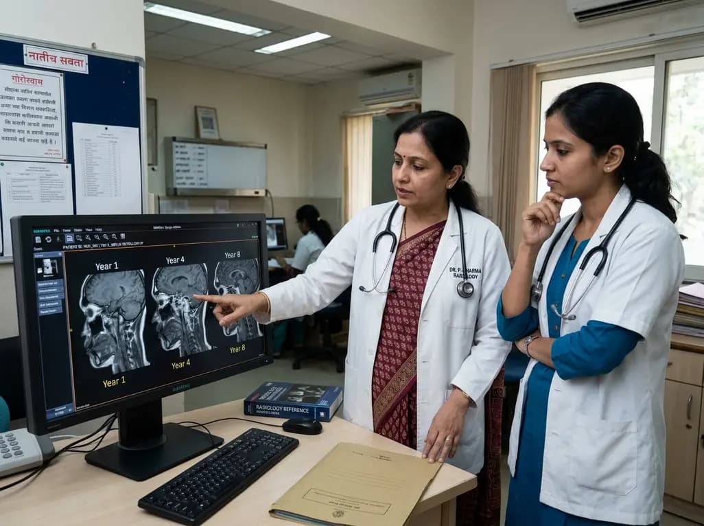
An eight-year longitudinal imaging follow-up reveals severe temporomandibular joint remodeling in nodulosis-arthropathy-osteolysis (NAO) syndrome. Despite extensive osseous destruction, the patient maintained functional jaw opening without TMJ interventions, highlighting the role of conservative management.
Today

Discover essential clinical insights into AGLP1-type amyloidosis, a rare localized complication of glucagon-like peptide-1 analog injections. Learn how histopathology and mass spectrometry differentiate benign iatrogenic amyloidomas from systemic disease, preventing misdiagnosis and unwarranted therapies.
Today
