.webp&w=384&q=75)

Loading, please wait...
Loading, please wait...
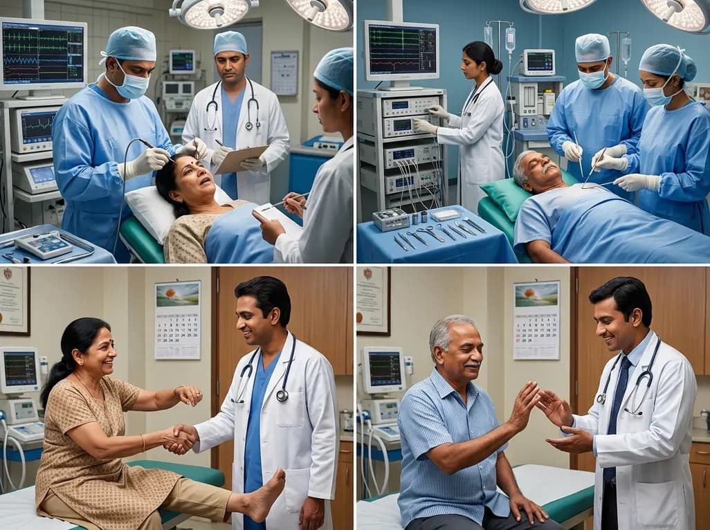
In the complex landscape of neuro-oncology, the surgical management of peri-rolandic gliomas represents one of the most challenging frontiers. Surgeons must navigate the delicate balance between achieving maximal safe resection and preserving motor function. Intraoperative motor mapping has emerged as the gold standard to achieve this objective. By identifying eloquent motor areas and subcortical tracts, clinicians can push the boundaries of resection without compromising the patient's quality of life. This article explores recent research comparing the efficacy of awake and asleep mapping techniques, specifically focusing on how these modalities impact outcomes across different molecular tumor subtypes. Historically, motor mapping was primarily performed in awake settings to allow for real-time functional assessment. However, advancements in neurophysiology have enabled robust motor mapping under general anesthesia. Consequently, neurosurgeons now have multiple strategies to manage tumors located in these critical regions. The goal remains consistent across all modalities: achieving the most extensive resection possible while maintaining the patient's motor integrity. As surgical technology continues to advance, the choice between awake and asleep mapping becomes increasingly nuanced. This evolution reflects a broader trend toward personalized surgical planning in oncology. Therefore, understanding the comparative outcomes of these techniques is essential for optimizing patient care in modern neurosurgery.
When evaluating intraoperative motor mapping, the choice between awake craniotomy and mapping under general anesthesia often depends on patient-specific factors. Recent clinical data suggest that both techniques demonstrate high efficacy in preserving neurological function. Specifically, researchers found that three-month postoperative deficit rates are remarkably comparable between the two cohorts. For instance, a study involving 130 patients showed that deficit rates were 5.6% for the awake group and 5.3% for the asleep group. This finding suggests that neither approach is inherently superior regarding long-term functional preservation. Furthermore, the selection of the mapping modality does not appear to adversely impact the primary safety outcomes. While awake mapping allows for direct communication with the patient, asleep mapping utilizes motor evoked potentials to monitor neural pathways. Notably, the technical success of these procedures is high in both settings. However, surgeons must account for the psychological and physiological demands of awake surgery. Some patients may not be suitable candidates for awake craniotomy due to anxiety or other medical contraindications. In contrast, asleep mapping provides a more controlled environment for the patient while still offering precise anatomical localization. Consequently, the decision-making process often involves a multidisciplinary approach involving neurosurgeons, anesthesiologists, and neurophysiologists. Ultimately, the parity in outcomes empowers surgical teams to select the modality that best suits the individual clinical scenario.
The impact of intraoperative motor mapping is also studied within the context of WHO 2021 molecular tumor subtypes. Specifically, the management of glioblastoma (GBM) and IDH-mutant gliomas may require different strategic focuses. In patients with IDH-mutant gliomas, who often have a longer life expectancy, preserving function over the long term is paramount. Conversely, in glioblastoma cases, the goal is often to balance radical resection with the preservation of performance status for subsequent therapies. Interestingly, recent studies indicate that the mapping modality choice does not significantly alter the functional outcomes for IDH-mutant patients. Moreover, the rates of new or worsening deficits remain low across these subtypes when mapping is utilized effectively. Additionally, the molecular characteristics of the tumor may influence the growth pattern and the degree of eloquence involvement. For example, some low-grade gliomas may exhibit more infiltrative growth into motor pathways compared to more circumscribed high-grade lesions. Therefore, the precision offered by cortical and subcortical mapping is vital regardless of the tumor's grade. By tailoring the surgical approach to the molecular profile, surgeons can better predict the disease course and set realistic functional goals. This integrated approach ensures that the surgical intervention aligns with the overall oncological management plan. Thus, molecular stratification remains a key component of modern neurosurgical strategy.
In the treatment of glioblastoma, intraoperative motor mapping plays a critical role in maintaining the Karnofsky Performance Status (KPS). Maintaining a high KPS is essential because it determines whether a patient can tolerate aggressive adjuvant chemoradiotherapy. Recent data reveals that patients undergoing surgery under general anesthesia often have significantly lower pre- and postoperative KPS scores compared to those in the awake group. However, this difference likely reflects selection bias, where sicker or more symptomatic patients are directed toward asleep procedures. Despite these baseline disparities, the mapping modality does not seem to influence overall survival in glioblastoma patients. Specifically, median survival times were comparable between the awake and asleep mapping groups. This suggests that as long as mapping is performed to facilitate safe resection, the choice of anesthesia does not dictate the oncological outcome. Furthermore, the length of hospital stay and discharge disposition were similar across both mapping cohorts. These findings are reassuring for clinicians who may prefer asleep mapping for patients with high-risk glioblastomas. Moreover, the ability to achieve a gross total resection remains the strongest predictor of survival. Consequently, the mapping technique should be viewed as a tool to reach that goal safely. By focusing on preserving function, surgeons ensure that patients remain eligible for the full spectrum of postoperative care.
Within the realm of asleep intraoperative motor mapping, different technical paradigms exist for stimulating the motor cortex. The two most common methods involve handheld probe direct cortical stimulation (HHP-DCS) and subdural electrode stimulation (SDE-DCS). Handheld probes allow for a dynamic and localized exploration of the cortical surface and subcortical pathways. In contrast, subdural electrodes provide the advantage of continuous monitoring throughout the resection phase. Recent comparative analyses indicate that both technical approaches result in similar rates of neurological deficits. Specifically, patients managed with HHP-DCS and SDE-DCS showed no significant difference in the frequency of postoperative motor decline. This parity suggests that the choice between these tools can be guided by surgeon preference and institutional resources. Additionally, the use of subcortical mapping is vital in both techniques to identify the proximity of the corticospinal tract. Therefore, regardless of the tool used, the focus must remain on the three-dimensional localization of motor fibers. Furthermore, some surgeons advocate for a combination of these methods to maximize safety. Notably, the integration of neuronavigation further enhances the accuracy of these mapping tools. As technology evolves, we may see further refinements in electrode design and stimulation protocols. Nevertheless, the current evidence supports the efficacy of both handheld and continuous monitoring strategies in the asleep setting.
Successfully implementing intraoperative motor mapping requires a streamlined surgical workflow and close collaboration. The surgical team must coordinate with neurophysiologists to ensure high-quality signal acquisition and interpretation. Furthermore, the anesthetic management is crucial, especially in asleep mapping where muscle relaxants must be avoided to obtain motor responses. In the awake setting, the transition from sedation to the testing phase must be handled with precision to avoid patient distress. Consequently, institutional experience plays a significant role in the safety and efficiency of these procedures. Training and standardized protocols can help minimize complications and improve the reliability of the mapping data. Moreover, preoperative counseling is essential to prepare patients for the unique environment of an awake craniotomy. By managing expectations and providing psychological support, teams can enhance the success rate of awake procedures. Additionally, postoperative rehabilitation should be integrated early for patients who experience transient deficits. This holistic approach ensures that the benefits of the surgery are maximized while minimizing the impact of potential complications. Ultimately, the successful application of motor mapping technology depends as much on the team's expertise as it does on the equipment itself. Through meticulous planning and execution, neurosurgeons can continue to improve outcomes for patients with peri-rolandic gliomas.
The primary goal involves achieving a maximal safe resection of the tumor while preserving critical neurological functions. By utilizing intraoperative motor mapping, neurosurgeons can accurately identify the primary motor cortex and descending tracts. This approach minimizes the risk of permanent motor deficits, thereby improving the patient's postoperative quality of life and performance status.
Current research indicates that both awake and asleep mapping techniques provide comparable rates of neurological deficits at three months. However, glioblastoma patients in the asleep cohort often present with lower preoperative performance status. Despite these baseline differences, overall survival rates remain similar between the two mapping modalities, suggesting both are effective surgical tools.
Subdural electrode stimulation allows for continuous monitoring of motor evoked potentials during the resection process. While handheld probes offer precise mapping at specific intervals, subdural electrodes provide a real-time safety net. However, recent studies suggest that the choice between these techniques often depends on the specific surgical context and the surgeon's institutional preference.
Disclaimer: This content is for informational and educational purposes only. It does not constitute professional medical advice, diagnosis, or treatment. Always seek the advice of your physician or other qualified healthcare provider with any questions you may have regarding a medical condition. Refer to the latest local and national guidelines for clinical practice.
References

Read summarized clinical updates, watch expert medical content, and earn CME certifications right from your smartphone.
Open your smartphone camera to scan and install.





A comparative analysis of awake and asleep intraoperative motor mapping for resecting peri-rolandic gliomas shows similar 3-month neurological outcomes across tumor subtypes, including glioblastoma and IDH-mutant cases.
3 months ago
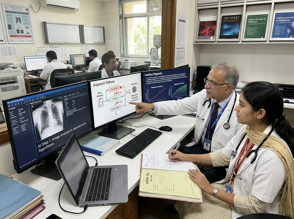
A breakthrough peer-reviewed study demonstrates that a sequential AI diagnosis system can mirror real-world clinical reasoning. By ordering stepwise tests and updating hypotheses dynamically, this multi-agent orchestrator achieved superior diagnostic accuracy while reducing investigative costs.
Today

A prenatal case highlights severe glutaric acidemia type II presenting as bilateral hyperechoic cystic kidneys at 18 weeks. Whole-exome sequencing revealed a novel homozygous ETFDH frameshift variant, demonstrating the power of rapid genetic testing in fetal renal anomalies.
Today

Review how tailored exercise programs benefit frail older patients with heart failure. Explore pathophysiological mechanisms, individualized training prescriptions, and strategies to overcome adherence barriers in clinical practice.
Yesterday

This comparative study evaluates posterior labral tears with and without glenohumeral instability, highlighting distinct demographic patterns, MRI underestimation in stable shoulders, and successful arthroscopic repair outcomes.
Yesterday

A clinical study demonstrates that early intervention with tocilizumab, either as monotherapy or bridged with corticosteroids, achieves significant proptosis reduction and resolves active inflammation in treatment-naive thyroid eye disease.
Yesterday
