.webp&w=384&q=75)

Loading, please wait...
Loading, please wait...

Diffuse gliomas present a formidable surgical challenge due to their infiltrative nature and epileptogenic potential. Consequently, modern neurosurgical oncology aims for maximal tumor resection while rigorously preserving functional neurological networks. Clinicians increasingly recognize that intraoperative electrocorticography offers powerful real-time electrophysiological insights during awake craniotomy. Traditional neuronavigation tools frequently fail to delineate microscopic boundaries between pathological tissue and functional brain parenchyma. Therefore, neurosurgeons require functional guidance to assess local cortex connectivity dynamically. Recent clinical investigations indicate that direct surface recordings expose critical differences between neoplastic masses and surrounding parenchyma. Furthermore, this technique captures spontaneous oscillatory rhythms directly from the exposed cerebral cortex. In addition, intraoperative recordings allow specialists to document functional cortical shifts in awake, responsive patients. As a result, neurosurgeons can adapt their surgical strategy before making irreversible parenchymal incisions. Thus, evaluating continuous electrical activity helps neurosurgeons navigate complex neurovascular anatomy safely. Notably, this functional assessment protects critical cortical pathways from accidental surgical disruption.
Direct recordings from the glioma mass expose severe disruption of normal neuroelectric architecture. Specifically, tumoral electrode contacts reveal a profound suppression across almost all physiological frequency bands. Alpha, beta, and gamma oscillations diminish markedly within the core neoplastic tissue. This widespread spectral attenuation reflects the progressive destruction of organized laminar cytoarchitecture and synaptic networks. In contrast, slow delta frequency activity displays a conspicuous and significant increase over tumoral regions. Pathologists and neurophysiologists associate this pathological delta slowing with regional tissue de-efferentation and local metabolic derangement. Moreover, researchers found that the severity of rhythmic loss directly mirrors the degree of neoplastic cellular infiltration. Even low levels of cortical tumor invasion reliably attenuate the brain's ability to generate physiological rhythms. Therefore, real-time spectral power mapping provides clinicians with immediate objective confirmation of malignant tissue replacement. Additionally, these spectral signatures remain distinct regardless of transient intraoperative anesthetic fluctuations. Consequently, the surgical team maintains continuous insight into tissue viability throughout the resection.
The brain tissue immediately surrounding a glioma represents an intricate microenvironment where neoplastic cells mingle with functional neurons. Consequently, delineating this transitional peritumoral cortex remains one of the most critical steps in modern neuro-oncology. Electrophysiological investigations demonstrate that the peritumoral zone exhibits a highly specific neuroelectric profile distinct from both tumor and healthy brain. Notably, peritumoral cortex displays a significant increase in relative beta frequency power. Furthermore, spectral analyses highlight marked modifications in aperiodic components, particularly steeper power spectrum slopes between 20 and 40 Hz. These distinct spectral alterations reflect altered synaptic excitation-inhibition balance driven by subtle glioma infiltration and glutamate-mediated excitotoxicity. However, unlike the tumor core, peritumoral tissue frequently retains active neural connectivity and contributes to critical neurological functions. Resecting these transitional regions without precise functional boundaries risks causing permanent neurological deficits. Hence, identifying increased beta power and altered spectral slopes helps surgeons locate the invasive tumor front accurately. In addition, understanding these peritumoral rhythm changes enhances seizure management strategies in patients presenting with tumor-associated epilepsy. Thus, peritumoral electrophysiology informs both oncological resection and postoperative functional recovery.
Interpreting multifaceted electrophysiological signals during a tense surgical procedure demands significant clinical expertise. To streamline this process, biomedical engineers developed automated classifiers utilizing advanced neural network architectures. These machine learning models evaluate real-time spectral power densities alongside aperiodic offset and slope values. Remarkably, the artificial neural network accurately differentiates electrode recordings into tumoral, peritumoral, and healthy categories without manual intervention. Consequently, this computational framework provides intraoperative decision support within seconds of cortical surface contact. Furthermore, automated signal decoding removes subjective interpretation and standardizes neurophysiological evaluation across different surgical centers. Clinicians can immediately visualize electrophysiological boundaries projected onto neuro-navigation displays. In addition, machine learning algorithms continuously refine their diagnostic precision by processing extensive multicenter spectral datasets. Similarly, rapid machine learning classification helps surgical teams identify epileptogenic peritumoral zones without delaying the operative workflow. Thus, intelligent intraoperative mapping establishes a reproducible standard for guided oncological resections. Overall, algorithmic evaluation enhances intraoperative precision and diminishes cognitive burden for surgical teams.
Diffuse gliomas require surgical teams to achieve a delicate balance between maximum oncological resection and functional preservation. While direct cortical electrical stimulation remains the conventional standard for functional mapping, it provides limited information regarding tumor margins. In contrast, surface electrocorticography continuously tracks physiological rhythms without provoking unwanted intraoperative seizures. Therefore, combining direct cortical stimulation with passive spectral recording delivers a comprehensive assessment of cortical functionality and pathology. Surgeons can precisely determine where neoplastic cell density drops and where functionally intact cortex begins. Furthermore, studies confirm that maximizing resection of infiltrated peritumoral cortex significantly extends progression-free survival in glioma patients. Minimizing residual infiltrative tumor also reduces subsequent recurrence rates and lowers the burden of secondary glioblastoma transformation. Concurrently, protecting functional peritumoral networks preserves language, executive cognition, and motor performance. Additionally, neurosurgical institutions can leverage this cost-effective recording methodology without requiring proprietary high-cost consumables. Consequently, widespread adoption will elevate surgical precision and oncological survival across diverse healthcare settings. Ultimately, these intraoperative electrophysiological advances empower neurosurgeons to push the boundaries of safe oncological cytoreduction.
Intraoperative electrocorticography records distinct spectral signatures across brain regions. Tumoral tissue demonstrates a marked suppression of physiological oscillatory rhythms alongside elevated delta slow-wave activity. In contrast, the peritumoral cortex exhibits preserved background architecture with increased relative beta band power and modified spectral slopes, distinguishing infiltrated margins from necrotic tumor core.
Diffuse gliomas aggressively infiltrate surrounding neural parenchyma beyond visible magnetic resonance imaging margins. Because this infiltrated peritumoral zone retains functioning neuronal circuits, indiscriminate resection causes severe neurological deficits. Electrophysiological profiling helps neurosurgeons achieve maximal safe oncological cytoreduction while sparing critical eloquent networks and mitigating postoperative cognitive or motor impairments.
Yes, neural network classifiers can analyze real-time power spectrum features, including frequency band distributions and aperiodic slopes. By processing these electrophysiological characteristics, automated algorithms accurately categorize electrodes into tumoral, peritumoral, or healthy tissue. This computational assistance provides objective, rapid margin guidance to the surgical team without prolonging operative duration.
Disclaimer: This content is for informational and educational purposes only. It is not intended to be a substitute for professional medical advice, diagnosis, or treatment. Always seek the advice of your physician or other qualified health provider with any questions you may have regarding a medical condition. Never disregard professional medical advice or delay in seeking it because of something you have read. While we strive to ensure that the information provided is accurate and up to date, medical knowledge evolves rapidly, and clinical guidelines may change. The content should not be relied upon as complete or exhaustive. Mention of specific products, tests, or procedures does not imply endorsement or recommendation. Reliance on any information provided here is solely at your own risk. Refer to the latest local and national guidelines for clinical practice.
References

Read summarized clinical updates, watch expert medical content, and earn CME certifications right from your smartphone.
Open your smartphone camera to scan and install.





Intraoperative electrocorticography during awake glioma surgery distinguishes tumor core from infiltrative peritumoral cortex using spectral biomarkers and neural network classifiers, optimizing maximal safe resection.
Today

A recent Journal of Clinical Oncology study demonstrates that industry payments to oncologists directly sway prescribing behavior. Oncologists who received marketing transfers favored promoted therapies over non-promoted alternatives, highlighting critical ethical concerns for evidence-based cancer practice.
Today

A recent qualitative study examines how college students seek digital sexual health information under persistent uncertainty. Discover the concept of functional trust, the clinical hazards of search abandonment, and evidence-based approaches clinicians can adopt to guide youth toward verified reproductive healthcare.
Today
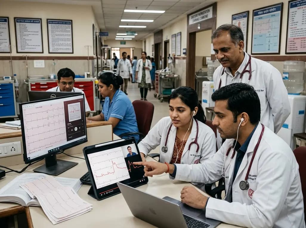
A randomized controlled trial demonstrates that curated FOAMed videos and podcasts produce superior gains in high-risk ECG interpretation and acute coronary syndrome clinical decision-making compared with print-based materials among interprofessional emergency care teams.
Today

A study reveals that Ginsenoside Rg3 attenuates osteoarthritis progression by modulating the Nrf2-mediated autophagy pathway, reducing chondrocyte apoptosis, and curbing extracellular matrix degradation, offering promising chondroprotective potential.
Today

A cross-sectional study demonstrates that lower pediatric motor competence strongly correlates with adverse metabolic syndrome markers, including central adiposity, elevated triglycerides, and low HDL cholesterol, emphasizing early neuromuscular interventions in youth with overweight and obesity.
Today
