.webp&w=384&q=75)

Loading, please wait...
Loading, please wait...
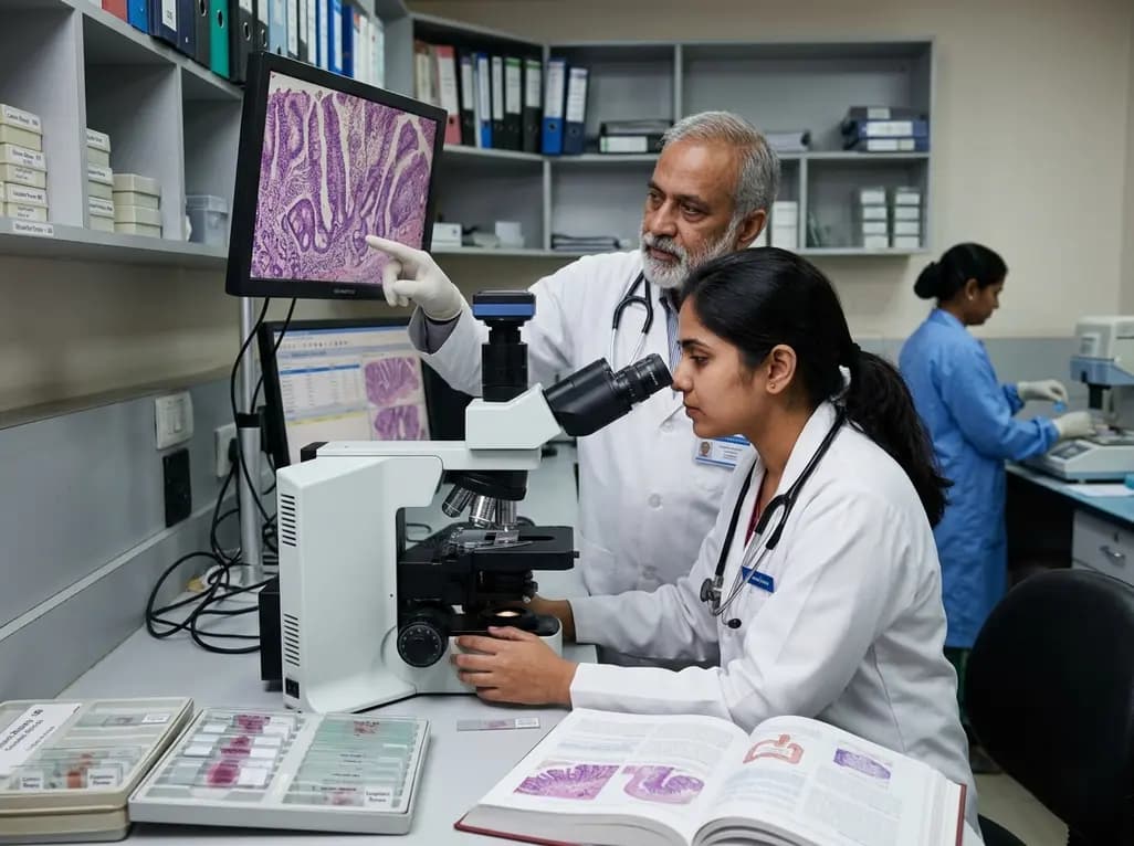
Colorectal cancer represents one of the most severe long-term complications in patients with long-standing colitis. Surveillance programs aim to detect precancerous changes before invasive carcinoma develops. However, evaluating IBD-associated colorectal dysplasia has become increasingly intricate. While classical lesions mirror sporadic adenomas, novel non-conventional morphologic variants pose unique diagnostic dilemmas for clinicians and pathologists alike. Recognizing these precursor lesions early remains vital to prevent malignant progression and guide clinical decision-making.
Historically, pathologists categorized dysplastic changes using the standard Riddell classification system. This traditional approach segregated findings into negative, indefinite, low-grade, or high-grade dysplasia. While this framework standardized surveillance for decades, it primarily captured conventional adenoma-like morphologies. Over time, clinicians encountered flat, invisible, or atypical lesions that failed to fit neatly into these conventional boxes.
Consequently, the medical community recognized significant diagnostic interobserver variability. Different institutions adopted divergent terminologies, which created substantial confusion in multicenter studies and routine patient care. To resolve these discrepancies, the 6th edition of the World Health Organization (WHO) Classification of Digestive System Tumours updated its criteria. This modernized framework formally integrates non-conventional dysplastic subtypes into mainstream pathology. Furthermore, it harmonizes histologic definitions across international platforms. As a result, gastroenterologists and pathologists can now communicate lesion risks with greater clarity and therapeutic precision.
Most surveillance biopsies demonstrate conventional dysplasia that features classic adenomatous architecture and pencil-shaped pseudostratified nuclei. Nevertheless, pathologists frequently encounter non-conventional variants with deceptive architectural patterns. These non-conventional entities include serrated lesions, hypermucinous dysplasia, goblet cell-deficient dysplasia, and crypt cell dysplasia.
Specifically, hypermucinous dysplasia displays prominent villous projections lined by tall cells containing abundant cytoplasmic mucin. Although these cells show minimal nuclear atypia, they carry a high risk of synchronous or metachronous invasive malignancy. Similarly, goblet cell-deficient dysplasia exhibits marked loss of mucin vacuoles accompanied by subtle nuclear enlargement. Serrated variants often resemble sporadic sessile serrated lesions or traditional serrated adenomas, yet they occur against a background of chronic mucosal inflammation. Because these variants present with subtle cytologic alterations, pathologists can easily misinterpret them as benign or reactive epithelium. Therefore, awareness of these distinct morphological variants is critical for accurate histological assessment.
Distinguishing genuine dysplasia from regenerative epithelial changes represents an ongoing diagnostic challenge. Chronic inflammation induces profound mucosal injury followed by active epithelial repair. During regeneration, epithelial cells develop enlarged nuclei, open chromatin, and prominent nucleoli that mimic true neoplastic transformation.
Moreover, active mucosal inflammation frequently obscures the boundary between regenerative atypia and low-grade dysplasia. In these ambiguous scenarios, pathologists often assign a diagnosis of "indefinite for dysplasia." To resolve difficult cases, immunohistochemical markers provide valuable objective assistance. For instance, aberrant p53 expression, such as complete absence or diffuse overexpression, strongly supports a diagnosis of genuine dysplasia. In contrast, patchy, weak p53 expression indicates reactive mucosal regeneration. Additionally, elevated Ki-67 proliferation extending to the mucosal surface confirms dysplastic growth. Consequently, combining immunohistochemistry with careful architectural analysis improves diagnostic confidence and reduces interobserver variation.
Accurate identification of precursor lesions directly dictates endoscopic and surgical interventions. Historically, any visible dysplastic lesion prompted immediate recommendation for total proctocolectomy. However, modern endoscopic advancements have radically shifted this paradigm toward organ-preserving therapies.
Currently, gastroenterologists utilize high-definition chromoendoscopy to delineate lesion borders with high precision. When an endoscopist identifies a discrete, well-circumscribed lesion in an uninflamed or quiescent segment, endoscopic resection is feasible. Techniques such as endoscopic mucosal resection (EMR) and endoscopic submucosal dissection (ESD) allow complete removal of polypoid or flat non-conventional lesions. Nevertheless, non-conventional variants often carry higher rates of multifocal disease within the same colonic segment. Therefore, complete endoscopic resection requires mandatory surrounding mucosal biopsies. If the pathologist identifies invisible dysplasia, multifocal high-grade dysplasia, or incomplete margins, surgical colectomy remains the definitive therapeutic choice.
Optimal patient care requires standardized pathology reporting that bridges microscopic findings and clinical interventions. Pathologists should explicitly describe the histologic subtype, the grade of dysplasia, and the status of resection margins. Furthermore, they should note any associated mucosal inflammation or architecture distortion in surrounding non-dysplastic tissues.
When a biopsy demonstrates partial features that do not meet definitive criteria for neoplasia, pathologists must clearly articulate the diagnostic uncertainty. Such cases benefit significantly from an expert gastrointestinal pathology second opinion. Moreover, regular multidisciplinary team meetings between gastroenterologists, pathologists, colorectal surgeons, and radiologists ensure tailored surveillance schedules. Patients harboring non-conventional dysplasia or persistent indefinite lesions require intensified endoscopic monitoring at shortened intervals. Ultimately, collaborative communication between disciplines prevents delayed cancer diagnoses and protects patients from premature, unnecessary radical bowel resections.
Understanding the molecular pathogenesis of colitis-associated carcinogenesis illuminates why non-conventional variants behave aggressively. Unlike sporadic colorectal cancer, which follows the traditional APC-initiated adenoma-carcinoma sequence, colitis-associated neoplasia frequently involves early TP53 mutations and widespread genomic instability.
Furthermore, chronic oxidative stress accelerates mucosal DNA damage across large continuous areas, creating extensive "field cancerization." This phenomenon explains why patients with non-conventional dysplasia frequently develop synchronous or metachronous neoplastic lesions elsewhere in the diseased colon. Emerging research focuses on next-generation sequencing and methylation profiling to identify high-risk molecular signatures before morphological dysplasia becomes conspicuous. Integrating molecular diagnostics with artificial intelligence-assisted endoscopy holds significant promise for future patient care. Consequently, clinicians will soon tailor surveillance intervals based on combined molecular risk profiles rather than endoscopic inspection alone.
Conventional dysplasia presents with typical adenomatous architecture, crypt crowding, and elongated, pseudostratified hyperchromatic nuclei that resemble sporadic adenomas. In contrast, non-conventional dysplasia encompasses distinct subtypes such as hypermucinous, goblet cell-deficient, and serrated variants. These lesions often display deceptively subtle cytologic atypia, architectural disarray, or flat morphology. Consequently, they are more difficult to identify endoscopically and histologically, yet they harbor a substantial risk for advanced colorectal neoplasia.
Immunohistochemical staining provides crucial objective evidence when distinguishing true dysplasia from severe reactive epithelial changes. Specifically, abnormal p53 expression, characterized by complete loss of staining or intense diffuse nuclear accumulation, strongly supports a neoplastic diagnosis. In contrast, reactive regenerative mucosa demonstrates patchy, wild-type p53 distribution. Additionally, aberrant topoisomerase II-alpha or expanded Ki-67 expression extending to the surface epithelium helps confirm true dysplastic proliferation in histologically equivocal cases.
Management depends heavily on lesion visibility, completeness of resection, and histological grade. Completely resected, discrete dysplastic lesions require close endoscopic surveillance with high-definition chromoendoscopy within three to six months. Conversely, finding invisible high-grade dysplasia, multifocal low-grade dysplasia, or unresectable non-conventional dysplasia warrants prompt surgical consultation for total proctocolectomy. Multidisciplinary teams must evaluate each patient individually to balance ongoing oncologic risks against the morbidity of radical surgical resection.
Disclaimer: This content is for informational and educational purposes only. It is not intended to provide medical advice or to be a substitute for professional medical assessment, diagnosis, or treatment. Patients should always seek the advice of a qualified physician or healthcare provider with any questions regarding a medical condition. Practitioners should rely on their own clinical judgement and expertise when evaluating patients and reviewing this information. Refer to the latest local and national guidelines for clinical practice.
References

Read summarized clinical updates, watch expert medical content, and earn CME certifications right from your smartphone.
Open your smartphone camera to scan and install.





A comprehensive overview of IBD-associated colorectal dysplasia, detailing the WHO 6th edition diagnostic classification, non-conventional morphologic variants, diagnostic pitfalls with regenerative atypia, and updated clinical management strategies.
4 weeks back

Targeting ferroptosis provides a compelling strategy to dismantle hypoxia- and acidosis-driven immunosuppression in solid tumors, enhancing response to modern immunotherapy.
Today

Findings from Project Viva indicate that sustained postpartum weight retention, rather than excessive gestational weight gain alone, directly worsens women's mid-life cardiovascular health, degrading blood pressure and glycemic parameters decades after delivery.
Today

A large UK Biobank study reveals that plasma proteomic signatures capture preclinical organ damage and improve multiorgan risk prediction across early CKM syndrome stages 0 to 2 beyond traditional PREVENT clinical models.
Today

A retrospective CBCT study reveals that superior TMJ space asymmetry correlates moderately with occipital condyle-atlas-axis asymmetry in unilateral TMJ osteoarthritis, highlighting an imaging correlate between jaw biomechanics and upper cervical alignment rather than a standalone diagnostic marker.
Today

A Bayesian multilevel meta-analysis reveals that aerobic training combined with moderate carbohydrate restriction modestly lowers HbA1c in type 2 diabetes. However, sparse data and very low certainty leave incremental benefits over exercise or diet alone unproven, highlighting the need for individualized care.
Today
