

Loading, please wait...
Loading, please wait...

Modern neuro-oncology faces an enduring clinical dilemma during the surgical management of isocitrate dehydrogenase (IDH)-wildtype glioblastoma. Standard surgical resections traditionally target tissue displaying gadolinium contrast enhancement on T1-weighted neuroimaging. However, contrast enhancement merely delineates areas of severe blood-brain barrier disruption. Consequently, conventional structural scans fail to capture the true anatomical boundary of invasive neoplastic cells. Highly aggressive glioma cells routinely infiltrate deep into surrounding non-enhancing brain parenchyma. Because fluid-attenuated inversion recovery (FLAIR) signal alterations combine infiltrative tumor with benign vasogenic edema, neurosurgeons cannot readily differentiate between them. Therefore, extending resection safely into non-enhancing parenchymal zones remains exceptionally difficult. In response to this diagnostic hurdle, investigators developed hypoxia-powered MRI to map microscopic metabolic alterations non-invasively. Glioblastoma cells adapt rapidly to microenvironmental oxygen deficits, driving pathological vascular remodeling and pronounced treatment resistance. In addition, hypoxic cellular niches shelter resilient glioma stem populations that trigger inevitable tumor recurrence. By mapping abnormal tissue oxygen kinetics rather than structural barrier breakdown, advanced physiological imaging addresses a critical neuro-oncological blind spot. Ultimately, precise biological localization empowers surgical teams to delineate invisible infiltrative margins with greater confidence and anatomical accuracy.
The clinical integration of functional neuroimaging requires both biological reliability and practical intraoperative usability. Hypoxia-powered MRI probes vascular physiology by employing blood oxygenation level-dependent (BOLD) sequences during transient, controlled hypoxia. During this non-invasive challenge, the patient breathes a brief gas mixture that mildly lowers arterial oxygen saturation. Deoxyhemoglobin serves as an endogenous paramagnetic contrast mechanism without requiring exogenous chemical contrast agents. Consequently, clinicians obtain dynamic physiological contrast that reflects regional microvascular responsiveness and oxygen extraction fraction. Furthermore, aberrant tumor neovasculature exhibits blunted or dysregulated hemodynamic responses compared to healthy cerebral tissue. Sophisticated computational algorithms process the resulting multi-temporal imaging data. Specifically, voxel-wise temporal decomposition isolates tissue response arrival, wash-in kinetics, and steady-state signal amplitude. Thereafter, automated Otsu thresholding identifies high-response hypoxic habitats located beyond the visible contrast-enhancing tumor perimeter. The surgical team then imports these coordinate-matched physiological habitat maps into standard neuronavigation software on the morning of surgery. Because image acquisition occurs the evening before intervention, the surgical schedule experiences minimal disruption. Thus, the technique transforms complex physiological measurements into actionable stereotactic targets suitable for real-time intraoperative guidance.
To validate this innovative strategy clinically, researchers launched the prospective GLIOXY initiative at University Hospital Zurich. The prospective investigation enrolled nine treatment-naive adult patients with histopathologically confirmed IDH-wildtype glioblastoma. The participant cohort included five men and four women with a median age of 62 years. Five patients presented with tumors localized to temporal or mesial temporal structures. On the evening prior to craniotomy, each participant completed the transient hypoxic BOLD MRI protocol under continuous physiological monitoring. Subsequently, computational pipelines generated normalized hypoxia response maps. The investigators then co-registered these response maps directly to anatomical structural MRI sequences. Furthermore, the neurosurgical team rigorously reviewed all proposed targets prior to skin incision. The surgeons carefully evaluated each potential biopsy site against eloquent cortical boundaries and major neurovascular structures. Consequently, only accessible, non-eloquent infiltrative zones received formal clearance for intraoperative biosurgical sampling. The neurosurgeon performed stereotactic sampling immediately following dural opening but before undertaking major cytoreductive tumor debulking. Finally, technicians immediately split each procured tissue core, allocating half for formalin-fixed paraffin embedding and freezing the remaining portion for molecular profiling.
The core clinical endpoint rested on confirming whether target habitats outside contrast enhancement genuinely harbored tumor cells. Across the cohort, neuronavigation transfer succeeded without error in all nine patients. In eight individuals, the surgical team confirmed favorable accessibility and successfully retrieved targeted tissue specimens. However, in one patient, vascular proximity rendered sampling unsafe, and the surgeon appropriately aborted target collection. Neuropathological evaluation of the eight retrieved specimens demonstrated infiltrative glioblastoma in seven cases. Therefore, the physiological targeting algorithm achieved an impressive positive predictive value of 87.5% in identifying neoplastic infiltration. Histological analysis revealed classical cellular features of malignant glioma, including cellular pleomorphism, nuclear atypia, and mitotic activity. In contrast, the single tumor-negative target revealed reactive astrocytosis and gliotic white matter without definitive tumor cells. Importantly, this discordant finding highlights the inherent complexity of differentiating reactive tissue inflammation from low-density infiltration. Nevertheless, identifying active neoplastic infiltration in seven out of eight prospective samples validates the physiological hypothesis. Thus, dynamic hypoxia mapping successfully distinguishes biologically active neoplastic infiltration from surrounding benign brain tissue far beyond conventional tumor borders.
Safety remains the paramount objective during any neurosurgical procedure, especially when extending resection boundaries toward functioning parenchyma. Crucially, the GLIOXY prospective protocol demonstrated complete biosurgical safety across all eight sampled patients. The surgical team recorded zero target-sampling complications, neurological deteriorations, or postoperative intracranial hemorrhages attributable to navigation-guided biopsies. Furthermore, operative durations increased only modestly because the team planned biopsy vectors before entering the operating room. This favorable safety profile supports the clinical viability of supramaximal resection strategies guided by functional physiology. Recent clinical trials suggest that extending cytoreduction into non-enhancing infiltrative margins improves both progression-free and overall patient survival. However, surgeons previously lacked objective physiological maps to differentiate salvageable functional cortex from infiltrated tissue. Conventional FLAIR hyperintensity includes extensive vasogenic edema, which often resolves after surgery or corticosteroid administration. In contrast, hypoxia-targeted BOLD imaging highlights metabolic disruption driven by viable infiltrative tumor cells. Consequently, this technology provides neurosurgeons with an objective, physiology-grounded roadmap to pursue safe supramaximal resection. By prioritizing functional preservation while expanding tumor clearance, clinicians can optimize biological cytoreduction without inflicting new functional neurological deficits.
Conventional neuroimaging relies on gadolinium contrast agents that identify blood-brain barrier disruption, which often misses infiltrative non-enhancing tumor cells. In contrast, hypoxia-powered MRI uses controlled transient hypoxia and endogenous deoxyhemoglobin to map microvascular physiological reactivity, identifying viable neoplastic niches in brain parenchyma that appear normal on contrast scans.
Glioblastoma cells routinely infiltrate beyond contrast-enhancing margins into adjacent brain parenchyma, causing nearly all localized recurrences. Targeting these non-enhancing invasive cells through supramaximal resection or focal therapy prolongs progression-free and overall survival. However, standard imaging cannot differentiate non-enhancing tumor invasion from benign vasogenic edema without functional metabolic mapping.
No surgical or neurological complications occurred among the eight sampled patients in the prospective trial. Neurosurgeons reviewed all candidate coordinates against anatomical eloquence and neurovascular structures prior to intervention. Sampling occurred safely via stereotactic neuronavigation immediately following dural opening, demonstrating that physiology-guided biopsies do not compromise intraoperative safety.
Disclaimer: This content is for informational and educational purposes only and is not intended to serve as medical advice. Healthcare professionals must exercise independent clinical judgment. While every effort has been made to verify data accuracy, the publisher and authors are not responsible for any clinical decisions made based on this material. Refer to the latest local and national guidelines for clinical practice.
References

Read summarized clinical updates, watch expert medical content, and earn CME certifications right from your smartphone.
Open your smartphone camera to scan and install.





The GLIOXY initiative demonstrates that hypoxia-powered BOLD MRI successfully identifies infiltrative tumor beyond standard contrast enhancement. Translating physiological hypoxia maps into neuronavigation enabled prospective stereotactic sampling with an 87.5% tumor yield and zero surgical complications.
Today

Maternal cardiovascular risk factors such as hypertension, obesity, and diabetes significantly elevate offspring cardiovascular disease risk through placental dysfunction and fetal programming. Enhancing preconception screening and antenatal care offers clinicians a powerful strategy to protect long-term heart health.
Today
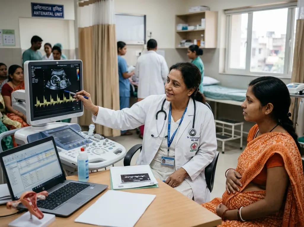
Non-RhD alloimmunization poses an emerging challenge in modern obstetric care as RhIG reduces RhD cases. This review highlights key non-RhD antigens, Doppler monitoring, and neonatal management strategies.
Today
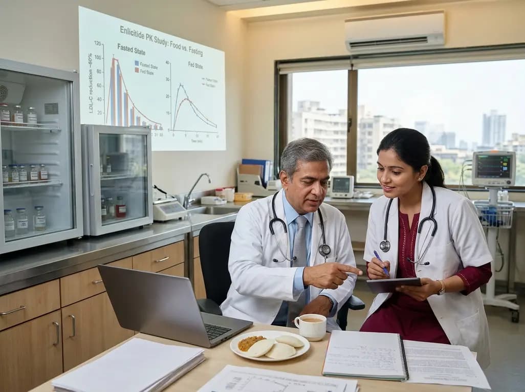
New phase I data evaluate the impact of food and beverages on enlicitide, the first oral PCSK9 inhibitor. While food reduces peak absorption, model simulations demonstrate robust LDL-C lowering of approximately 60%, confirming meaningful pharmacokinetic forgiveness and flexible real-world administration.
Today

A retrospective cohort study reveals that lumbar spine bone mineral density does not distinguish extremity long bone fracture status in adults with severe motor and intellectual disabilities. Because age drove fracture risk rather than spinal density, clinicians must adopt comprehensive assessment tools.
Today

A landmark UK Biobank study by Karolinska Institutet researchers reveals that optimal rest mitigates neurodegeneration in cardiometabolic illness. Patients maintaining high sleep quality demonstrated superior cognitive scores and larger brain volumes, highlighting sleep optimization as an actionable clinical strategy.
Yesterday
